Figures & data
Figure 6 Apoptosis and survival of GFP-MSCs at implantation site.
Notes: TUNEL analysis shows that apoptotic cells peaked at 1 week, and then decreased over time in animals grafted with labeled GFP-MSCs (A), unlabeled GFP-MSCs (B), and PBS (C). Graph show the percentages of TUNEL-positive cells in animals grafted with labeled and unlabeled GFP-MSCs (D). Graph shows the number of apoptotic GFP-MSCs in animals grafted with labeled and unlabeled GFP-MSCs (E). Graph shows the percentage of viable GFP-positive cells in animals grafted with labeled and unlabeled GFP-MSCs (F). *p<0.05 between labeled GFP-MSCs and PBS; #p<0.05 between unlabeled GFP-MSCs and PBS; Bar =15 μm.
Abbreviations: MSC, mesenchymal stem cell; PBS, phosphate-buffered saline.
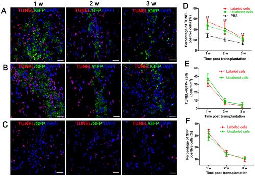
Figure 8 Histology of the grafted cells in the injection site.
Notes: Fluorescence immunostaining micrographs show that GFP-MSCs remained in the injection site in animals grafted with polymersome-labeled GFP-MSCs and unlabeled GFP-MSCs. Only a few surviving GFP-MSCs differentiated into GFAP-positive astrocytes (arrows), but not into NeuN-positive neurons. A large number of cells were phagocytized by macrophages (arrows) (A). Graphs show the numbers of GFAP+GFP+ (B), CD11b+GFP+ (C), and the percentage of GFP-MSCs (D) in animals grafted with polymersome-labeled GFP-MSCs and unlabeled GFP-MSCs. Bar =15 μm.
Abbreviation: MSC, mesenchymal stem cell.
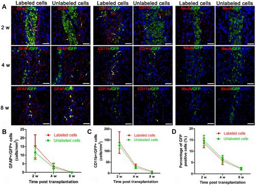
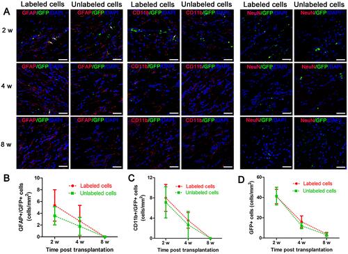
Figure 9 Histology of migrating cells in the corpus callosum.
Notes: Fluorescence immunostaining micrographs show that a small amount of GFP-MSCs migrated to the corpus callosum in animals treated with polymersome-labeled cells and unlabeled cells. Only a few surviving GFP-MSCs were differentiated into GFAP-positive astrocytes (arrows) and were phagocytized by macrophages (arrows), but no cells differentiated into NeuN-positive neurons (A). Graphs show the numbers of GFAP+GFP+ (B), CD11b+GFP+ (C), and GFP-MSCs (D) in animals grafted with polymersome-labeled GFP-MSCs and unlabeled GFP-MSCs. Bar =15 μm.
Abbreviation: MSC, mesenchymal stem cell.