Figures & data
Figure 1 Percentage of ibuprofen released from nanoparticles versus time.
Notes: In vitro release of ibuprofen from nanoparticles was assessed as described in the Materials and methods. Measurements were carried out every 30 minutes by means of UV-VIS spectroscopy with a Shimadzu UV mini-1240. The results are presented as the percentage of ibuprofen released over time. The experiment was performed in triplicate.
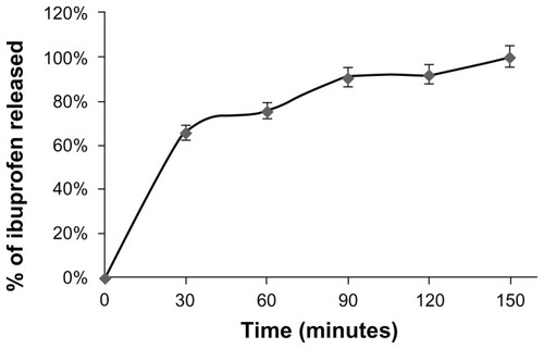
Figure 2 Effect of ibuprofen, PLGA NPs and ibuprofen-loaded PLGA NPs on cell proliferation.
Notes: MKN-45 cells (5 × 10⁴ cells/well) were seeded in 24-well plates and incubated overnight. The cells were then treated with ibuprofen, ibuprofen-loaded PLGA NPs, or PLGA NPs for 24 hours. The cells were harvested and counted. Viability was determined by Trypan blue dye exclusion. Cell growth was calculated by averaging cell counts and as expressed as the percentage of the control, represented by MKN-45 cells grown in culture medium alone or culture medium with DMSO (100%). The mean values from three independent experiments performed in duplicate are presented. Ibuprofen significantly inhibited the proliferation of MKN-45 cells only in cells treated with ibuprofen-loaded PLGA NPs. Bars, SD; *P < 0.5; †P < 0.005; Student’s t-test.
Abbreviations: PLGA, poly(lactic-co-glycolic acid); NPs, nanoparticles; DMSO, dimethyl sulfoxide.
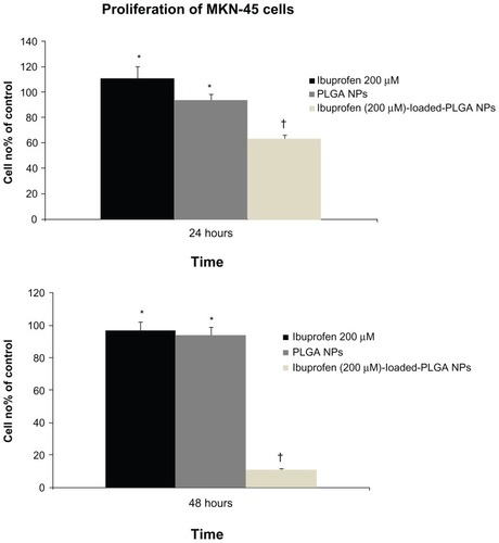
Table 1 Effect of serum-free conditions on cell cycle in MKN-45 cells
Figure 3 Nanoparticle uptake by MKN-45 cells revealed by fluorescence microscopy.
Notes: Intracellular detection of ibuprofen-loaded PLGA NPs. Cells were imaged using a Leica fluorescence microscope at 40 × objective magnification. A total of 2 × 10⁴ MKN-45 cells were seeded on Millicell EZ slides as described in the Materials and methods and treated with ibuprofen-loaded PLGA NPs for 2 hours. The nuclei were stained with DAPI. The localization of NPs was intracellular.
Abbreviations: PLGA, poly(lactic-co-glycolic acid); NPs, nanoparticles; DAPI, 4′,6-diamidino-2-phenylindole.
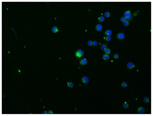
Figure 4 Nanoparticle uptake by MKN-45 cells revealed by flow cytometry. A total of 5 × 10⁴ MKN-45 cells were seeded in a 24-well plate for 24 hours prior to assay. The medium was then replaced with fresh RPMI 1640 without serum before ibuprofen-loaded PLGA NPs were added. After 2 hours of incubation, the cell monolayers were rinsed three times with PBS buffer to remove excess NPs and incubated with complete medium. NP uptake was verified by flow cytometry. The cells were collected and washed twice with cold PBS. The pellets were resuspended in FACSFlow Sheath Fluid and analyzed by a FACSCanto II cytometer. For each sample, >20,000 cells were analyzed by FACSDiva software. Typical fluorescence histograms (A) and the percentage of fluorescent cells (B) at different time intervals are shown. Nearly 100% of the cells were fluorescent at 24 and 48 hours.
Notes: Values represent the means of three independent experiments performed in triplicate; (†P < 0.005; Student’s t-test).
Abbreviations: PLGA, poly(lactic-co-glycolic acid); NPs, nanoparticles; PBS, phosphate-buffered saline.

Table 2 Release of ibuprofen from PLGA NPs in cells
Figure 5 Real-time PCR analysis.
Notes: Analysis of f ANGPTL4 mRNA levels in MKN-45 cells treated with free ibuprofen (200 and 800 μM) or ibuprofen-loaded NPs for 2 hours by Real-Time PCR. Each sample was normalized against human GAPDH. Bars indicate the fold change values versus untreated MKN-45 cells of three independent experiments performed in triplicate. Bars, SD; *P < 0.005; Student’s t-test.
Abbreviations: NPs, nanoparticles; GAPDH, human glyceraldehyde 3-phosphate dehydrogenase.
