Figures & data
Figure 1 Schematic illustrations of nanoparticles.
Notes: (A) Schematic illustrations for the synthesis of the FeDM and FeDM-FITC-SLN. (B) Schematic diagram for the absorption and transport of SLNs in different intestinal lesions.
Abbreviations: FeDM, Fe3+ and L-dopa methyl ester hydrochloride complexes; FITC, fluorescein isothiocyanate; SLN, solid lipid nanoparticle; ODA, octadecylamine.
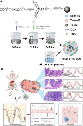
Figure 2 Characterization of nanoparticles.
Notes: (A and B) Transmission electron microscopy images of FeDM-FITC-SLNs. (C) Histogram of nanoparticle size distribution. (D) T1-weighted MR images of FeDM and FeDM-FITC-SLNs solutions at different Fe concentration. (E) Linear fitting of the inverse T1 values.
Abbreviations: FeDM, Fe3+ and L-dopa methyl ester hydrochloride complexes; FITC, fluorescein isothiocyanate; SLN, solid lipid nanoparticle.
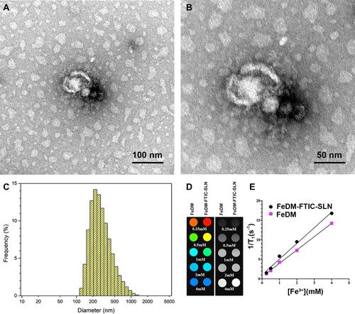
Figure 3 Toxicity assay of nanoparticles.
Notes: (A) CT-26 cells in 96-well plates were treated with different concentrations of FeDM or FeDM-FITC-SLN for different times; cell viability data was determined using the CCK-8 assay. (B) After 14 days, colon (I and IV), kidney (II and V), and liver (III and VI) were harvested and taken for histopathology. Control (I, II, and III) and experimental (IV, V, and VI) animals were included.
Abbreviations: FeDM, Fe3+ and L-dopa methyl ester hydrochloride complexes; FITC, fluorescein isothiocyanate; SLN, solid lipid nanoparticle.
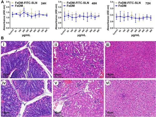
Table 1 Mice Were Observed Daily for Changes in Body Weight for 14 Days (Day 1 to Day 14)
Table 2 Effect on Serum Chemistry Profiles After Administration
Table 3 MR Signal-to-Noise Ratio (SNR) for Intestine in the Different Mouse Models
Figure 4 Cellular uptake of nanoparticles in vitro.
Notes: Cells were incubated with 800 μg/mL FeDM-FITC-SLNs at 37 °C for 24 h and then stained with Hoechst and LysoTracker Red. Images were captured using confocal microscopy. Blue represents nuclei stained with Hoechst dye; red represents lysosome; green represents FITC-labeled SLNs; and yellow shows colocalization of SLNs and lysosomes. (A) magnification, ×100; (B) magnification, ×200; and (C) magnification, ×600.
Abbreviations: FITC, fluorescein isothiocyanate; SLN, solid lipid nanoparticle.
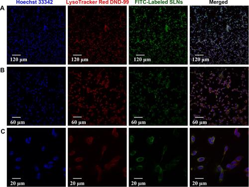
Figure 5 In vivo MR imaging.
Notes: In vivo T1-weighted MRI of the experimental mouse models before and after intravenous injection of Gd-DTPA (A) and enema of FeDM-FITC-SLN (B). The white arrows indicate intestines.
Abbreviations: Gd-DTPA, gadolinium-diethylenetriaminepentaacetic acid; FeDM, Fe3+ and L-dopa methyl ester hydrochloride complexes; FITC, fluorescein isothiocyanate; SLN, solid lipid nanoparticle; AUC, acute ulcerative colitis; CUC, chronic ulcerative colitis; COAD, colon adenocarcinoma.
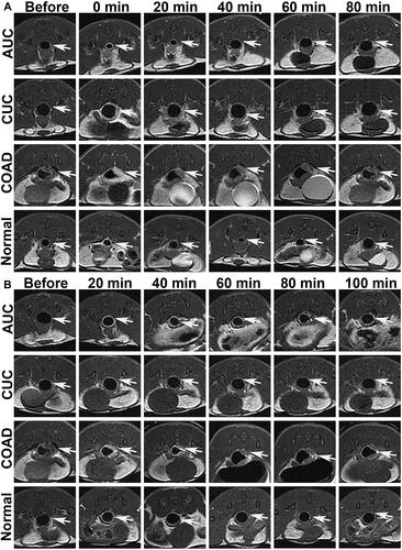
Figure 6 Imaging analysis for MRI.
Notes: MR SNR for the intestine in different mouse models ((A) acute ulcerative colitis; (B) chronic ulcerative colitis; (C) colon adenocarcinoma; (D) normal) after intravenous injection of Gd-DTPA and enema of FeDM-FITC-SLN. *p < 0.05 vs before; **p < 0.01 vs before; ***p < 0.001 vs before.
Abbreviations: SNR, signal-to-noise ratio; Gd-DTPA, gadolinium-diethylenetriaminepentaacetic acid; FeDM, Fe3+ and L-dopa methyl ester hydrochloride complexes; FITC, fluorescein isothiocyanate; SLN, solid lipid nanoparticle; AUC, acute ulcerative colitis; CUC, chronic ulcerative colitis; COAD, colon adenocarcinoma.
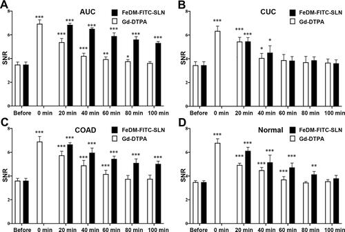
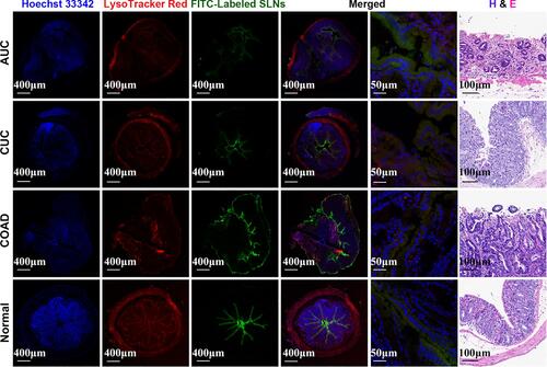
Figure 7 Histologic analysis after MR imaging.
Notes: Confocal laser scanning microscopy images and hematoxylin and eosin-stained images of intestinal issues in different mouse models after enema of FeDM-FITC-SLNs. Yellow is colocalization of SLNs and intestinal issues.
Abbreviations: H&E, hematoxylin-eosin staining; FITC, fluorescein isothiocyanate; AUC, acute ulcerative colitis; CUC, chronic ulcerative colitis; COAD, colon adenocarcinoma.