Figures & data
Table 1 Relationship Between the Expression of HOTAIR and Clinicopathological Features in Endometriosis
Figure 1 HOTAIR expression is upregulated and miR-761 expression is downregulated in ectopic endometrial tissues. (A) Expression of HOTAIR in normal endometrium, eutopic endometrium, and ectopic endometrium was detected by RT-qPCR. (B) miR-761 expression in different types of endometrium was detected by RT-qPCR. (C) Correlation between the expression of HOTAIR and miR-761 in ectopic endometrium analyzed by Pearson’s correlation analysis. **P < 0.01.
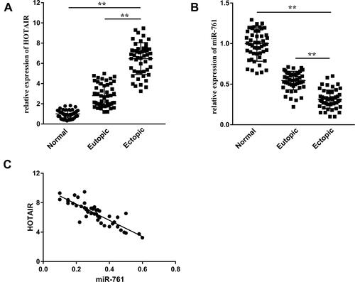
Figure 2 ESCs transport HOTAIR to surrounding cells through exosomes. (A) Exosome morphology as observed by TEM. Scale bar = 100 nm. (B) NTA was employed to detect the size and concentration of exosomes. (C) Expression of two biomarkers of exosomes, CD9 and CD63, was measured by Western blotting. (D) Internalization of DiI-labeled exosomes (red) in DAPI-labeled ESCs (blue). Scale bar = 20 μm. (E) HOTAIR expression in exosi-HOTAIR, exosi-HOTAIR-control, exoOe-HOTAIR, and exoOe-control. (F) HOTAIR expression in ESCs co-cultured with different types of exosomes. **P < 0.01.
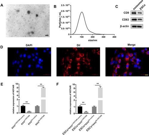
Figure 3 Exosomal HOTAIR promotes the proliferation, migration, and invasion, and inhibits the apoptosis of ESCs. (A) The CCK-8 assay was used to evaluate ESCs proliferation. (B) ESCs proliferation was examined by EdU assay. (C) The migration and invasion of ESCs were detected by transwell assays. (D) Flow cytometry was applied to detect ESCs apoptosis. (E) The tube formation capacity of HUVECs treated with different exosomes was determined. **P < 0.01.
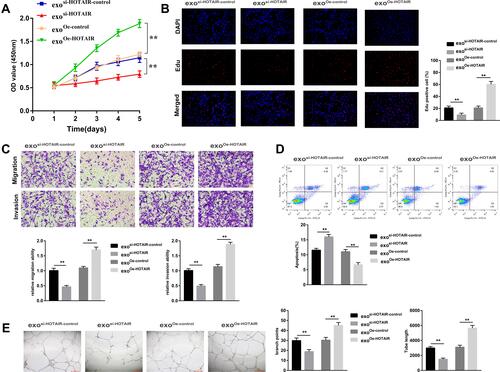
Figure 4 HOTAIR acts as a ceRNA to react with miR-761 to regulate HDAC1. (A) Binding sites and mutant sites between miR-761 and HOTAIR. (B) The dual-luciferase reporter assay was used to detect the combination of HOTAIR and miR-761. (C) The combination of HOTAIR and Ago2 was detected by the RIP assay. (D) Enrichment of HOTAIR expression by miR-761 was detected by the RNA pull-down assay. (E) Binding sites between miR-761 and HDAC1. (F) The binding between miR-761 and MDM2 was assessed by the dual-luciferase reporter assay. (G) Protein expression of HDAC1 was detected by Western blotting. **P < 0.01.
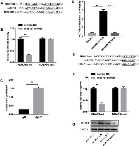
Figure 5 Exosomal HOTAIR promotes the proliferation, migration, and invasion of ESCs and angiogenesis of HUVECs, and inhibits ESCs apoptosis through sponging miR-761. (A) miR-761 expression measured by RT-qPCR. (B) The CCK-8 assay was used to evaluate ESCs proliferation. (C) ESCs proliferation was examined by the EdU assay. (D) The migration and invasion of ESCs were detected by transwell assays. (E) Flow cytometry was applied to detect ESCs apoptosis. (F) The angiogenesis capacity of HUVECs was evaluated by the tube formation assay. **P < 0.01.
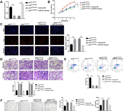
Figure 6 Exosomal HOTAIR promotes the proliferation, migration, and invasion of ESCs and angiogenesis of HUVECs, and inhibits ESCs apoptosis through upregulating HDAC1. (A) mRNA expression of HDAC1 measured by RT-qPCR. (B) Protein expression of HDAC1 measured by Western blotting. (C) The CCK-8 assay was used to evaluate ESCs proliferation. (D) ESCs proliferation was examined by the EdU assay. (E) The migration and invasion of ESCs were detected by transwell assays. (F) Flow cytometry was applied to detect ESCs apoptosis. (G) The angiogenesis capacity of HUVECs was evaluated by the tube formation assay. **P < 0.01.
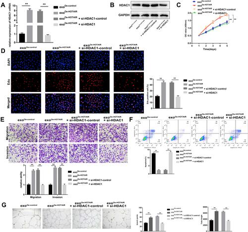
Figure 7 The HOTAIR/miR-761/HDAC1 axis activates STAT3-mediated inflammation. (A) Protein expression of STAT3 and p-STAT3 was measured. (B) Expression of STAT3 and p-STAT3 in ESCs co-transfected with HOTAIR or miR-761 mimics. (C) Cell proliferation was detected using the CCK-8 assay. (D) Cell migration and invasion was evaluated using transwell assays. (E) The angiogenesis capacity of HUVECs was evaluated by the tube formation assay. (F) ELISA was used to measure the levels of IL-1β, IL-6, IL-8, and TNF-α. **P < 0.01.
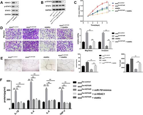
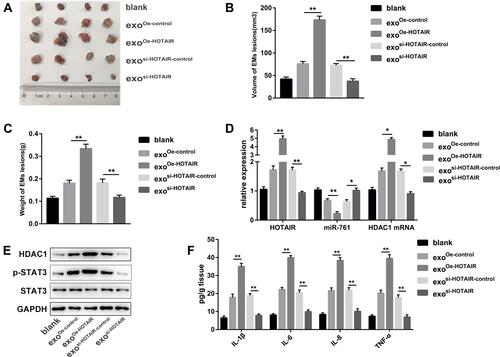
Figure 8 Exosomal HOTAIR promotes the growth of endometrial lesions in vivo. (A) Photograph of endometrial lesions. (B) Total volume of endometrial lesions. (C) Total weight of endometrial lesions. (D) RT-qPCR was carried out to measure expression of HOTAIR, miR-761, and HDAC1 mRNA in different group. (E) Protein expression of HDAC1, STAT3, and p-STAT3 was detected via Western blotting. (F) ELISA was used to measure the levels of IL-1β, IL-6, IL-8, and TNF-α in endometrial lesions. *P < 0.05, **P < 0.01.