Figures & data
Table 1 Overview of nanoparticle (NP) formulations in development for imaging and drug delivery with examples for approved drugs, with indication of the most important fields of applicationCitation172
Figure 1 Targets for cytotoxicity of nanoparticles (NPs).
Notes: NPs may act through extracellular generation of reactive oxygen species (ROS) (1), they may physically damage the plasma membrane by causing holes (2) or bind to membrane proteins like nicotinamide adenine dinucleotide phosphate-oxidase (3), Ca2+ channels (4), and membrane receptors (5), thereby inducing oxidative signaling, increasing intracellular Ca2+ levels and activating second-messenger cascades. Inside the cells, NPs may interfere with mitochondrial metabolism (6), causing generation of radicals and induction of apoptosis. Intracellular ROS generation by NPs or by metals from lysosomal degradation (7) as well as lysosomal disruption (8) and direct binding to components of the cytoskeleton (9) and the induction of structural alterations of proteins (10) are additional modes of toxic actions. In the nucleus, interference with the transcription machinery and oxidative damage of the DNA (11) may occur.
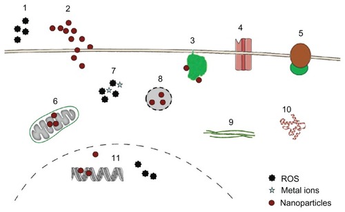
Figure 2 Simplified representation of active uptake mechanisms in nonphagocytic cells.
Notes: Nanoparticle (●) uptake has been evaluated mainly according to macropinocytosis, represented here as only one route, through macropinosome (MP), clathrin-mediated uptake by clathrin-coated pits (CC), and caveolae-dependent uptake by caveosomes (Cav). Uptake by clathrin-independent caveolae-independent endocytosis, which includes flotillin-, Arf6-, Cdc42-, and RhoA-dependent uptake, is also presented as only one route. Fluid-phase endocytosis, which mainly uses the clathrin-coated pits, is not depicted as a separate route. All pathways deliver their content to endosomes (E), late endosomes (LE) and lysosomes (L); the content of caveolosomes may also be delivered to the endoplasmic reticulum (ER) and the Golgi apparatus. Vesicular transport through the cell occurs through transcytotic vesicles (TV). © 2012, Elsevier. Reproduced with permission from Fröhlich E, Roblegg E. Models for oral uptake of nanoparticles in consumer products. Toxicology. 2012;291(1–3):8.Citation179

Table 2 Routes of endocytic uptake in nonphagocytic cells