Figures & data
Table 1 Physicochemical properties of drug-free and drug-loaded micelles
Figure 1 Synthetic scheme of NCHGC.
Abbreviations: EDC, 1-ethyl-3-(3-dimethylaminopropyl)-carbodiimide hydrochloride; CHGC, cholesterol-modified glycol chitosan; SMCC, N-succinimidyl 4-(maleimidomethyl) cyclohexane carboxylate; NCHGC, nuclear localization signal-conjugated cholesterol-modified glycol chitosan; NLS, nuclear localization signal.
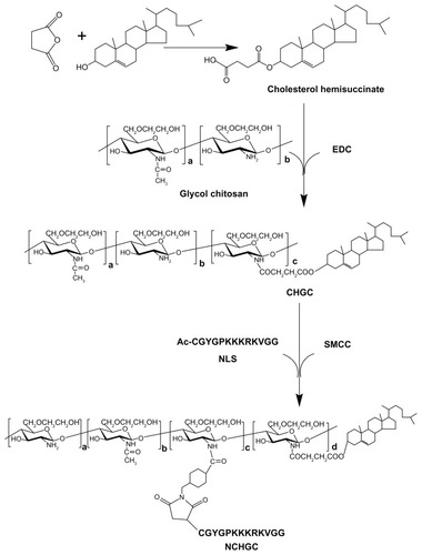
Figure 2 Proton nuclear magnetic resonance spectra of (A) glycol chitosan, (B) cholesterol-modified glycol chitosan (CHGC), (C) nuclear localization signal (NLS) (Ac-CGYGPKKKRKVGG), and (D) NLS-conjugated cholesterol-modified glycol chitosan (NCHGC).
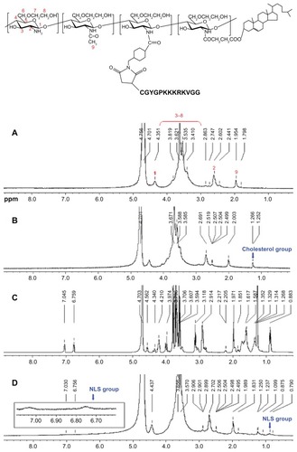
Figure 3 Plots of the intensity ratio I338/I333 vs log C for nuclear localization signal-conjugated cholesterol-modified glycol chitosan with various compositions.
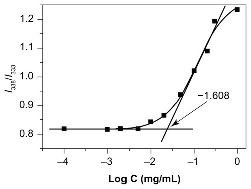
Figure 4 Schematic illustration of the preparation procedure of doxorubicin-loaded nuclear localization signal-conjugated cholesterol-modified glycol chitosan micelles by an emulsion/solvent evaporation method.
Abbreviation: NLS, nuclear localization signal (Ac-CGYGPKKKRKVGG).
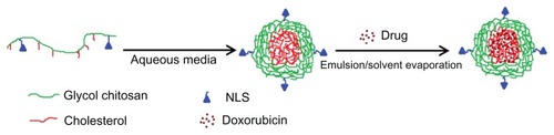
Figure 5 Transmission electron microscopy image of doxorubicin-loaded nuclear localization signal-conjugated cholesterol-modified glycol chitosan micelles (×20,000).
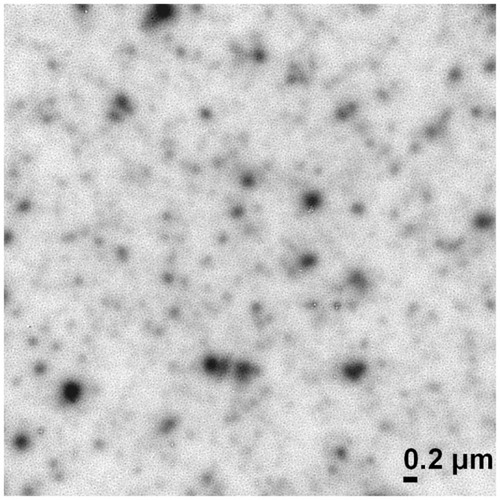
Figure 6 Release profiles of doxorubicin (DOX) from doxorubicin-loaded cholesterol-modified glycol chitosan (DCHGC) and doxorubicin-loaded nuclear localization signal-conjugated cholesterol-modified glycol chitosan (DNCHGC) micelles at 37°C in phosphate-buffered saline containing 10% fetal bovine serum at pH 7.4.
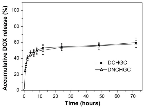
Figure 7 Flow cytometric analysis of HeLa cells incubated with free doxorubicin (DOX), doxorubicin-loaded cholesterol-modified glycol chitosan (DCHGC) and doxorubicin-loaded nuclear localization signal-conjugated cholesterol-modified glycol chitosan (DNCHGC) micelles for 2 hours.
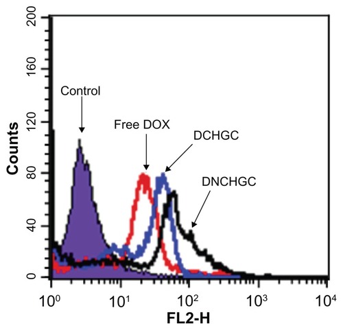
Figure 8 Confocal laser scanning microscopy images of HeLa cells after incubation with (A) free doxorubicin (DOX), (B) doxorubicin-loaded cholesterol-modified glycol chitosan micelles and (C) doxorubicin-loaded nuclear localization signal-conjugated cholesterol-modified glycol chitosan micelles for 2 hours. The red DOX fluorescence was observed in (A1–C1). The nuclei were stained with Hoechst 33342 in (A2–C2), and the merged photos were presented in (A3–C3).
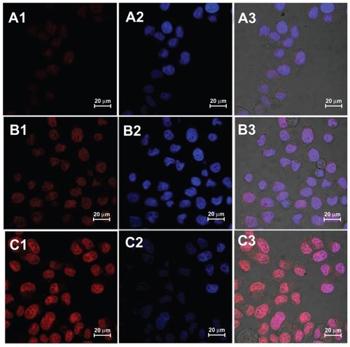
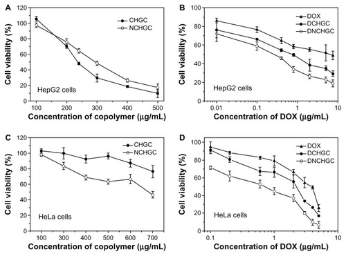
Figure 9 In vitro cytotoxicity of blank micelles and various doxorubicin (DOX) formulations against HepG2 and HeLa cells after 48 hours of incubation. (A) Cholesterol-modified glycol chitosan (CHGC) and nuclear localization signal-conjugated CHGC (NCHGC) micelles in HepG2 cells; (B) free DOX, doxorubicin-loaded CHGC (DCHGC) and doxorubicin-loaded nuclear localization signal-conjugated CHGC (DNCHGC) micelles in HepG2 cells; (C) CHGC and NCHGC micelles in HeLa cells; and (D) free DOX, DCHGC and DNCHGC micelles in HeLa cells.