Figures & data
Figure 1 The schematic diagram and feature of the synthesis of Lec(RSV). (A) Schematic representation of the utilization of lecithin and resveratrol to form Lec(RSV) nanoparticles, (B) nanoprecipitation method was used to achieve soluble Lec(RSV).
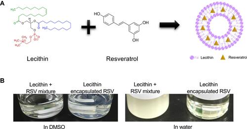
Table 1 The Average Size of Nanoparticles
Figure 2 The size of empty Lec nanoparticle and Lec(RSV) nanoparticle. (A) TEM analysis of empty Lec nanoparticle. (B) TEM analysis of Lec(RSV) nanoparticles; Orange scale bar indicated 200 nm; while scale bar indicated 100 nm. (C) The plot of the average hydrodynamic size of Lec(RSV) nanoparticles; (D) the plot of zeta potential of Lec(RSV) nanoparticles.
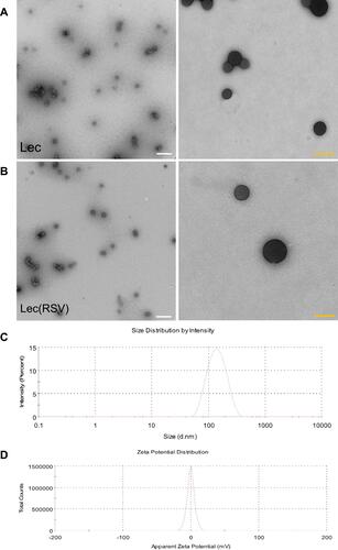
Table 2 The Encapsulation Efficacy of RSV
Figure 3 The stability of Lec and Lec(RSV) nanoparticles. (A) The size of Lec and Lec(RSV) nanoparticles during a 48-hour period; (B) the size of Lec(RSV) nanoparticles at predetermined time points (day-1, 4, 8, 15, 30, and 60); (C) the appearance of Lec(RSV) after long-term storage of 12 months; (D) the release of RSV of Lec(RSV) nanoparticles after encapsulation.
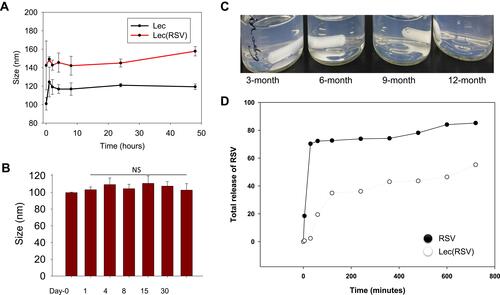
Figure 4 The in vitro uptake ability and cytotoxicity of Lec and Lec(RSV) nanoparticles. (A and B) The BT474 breast cancer cells uptake of Lec and Lec(RSV) encapsulated FITC; (C) in vitro cytotoxicity assay to observe the cancer cell killing ability of Lec, RSV, and Lec(RSV), respectively; (D) ROS scavenging effects of Lec, RSV and Lec(RSV). ***Indicated significance value of p < 0.001; compared to control.
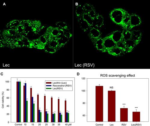
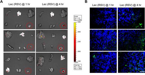
Figure 5 The in vivo tumoral uptake of Lec(RSV) nanoparticle. (A) BT474 orthotopic model was used to carry out in vivo tumoral uptake experiment. We treated one group of female Balb/C nude mice (n = 3) with Lec(RSV + FITC) NPs for 1 hour and another group (n = 3) for 4 hours. The red circles indicate the accumulation effects of the nanoparticles in the tumor at 1 hour and 4 hours after injection. It shows that the tumoral uptake of Lec(RSV) was significantly increased in the tumor after 4 hours compared to the 1-hour group, indicating the reliability of Lec(RSV) to be used as a drug delivery system for the treatment of cancer. (B) TUNEL assay analysis of the tumor section in (A). At 4 hours, the tumors treated with Lec(RSV) indicated higher apoptosis level as compared to the tumors treated only for 1 hour, indicated prolonged effect of tumor killing effect of Lec(RSV). Scale bar indicated 50 µm.