Figures & data
Figure 1 Curcuma longa plant (A), C. longa tubers (B), and C. longa powder (C).
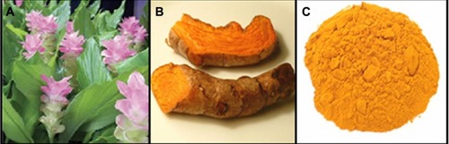
Figure 2 Photograph of Curcuma longa (A) and silver/C. longa (B) emulsions after 24 hours of stirring time.
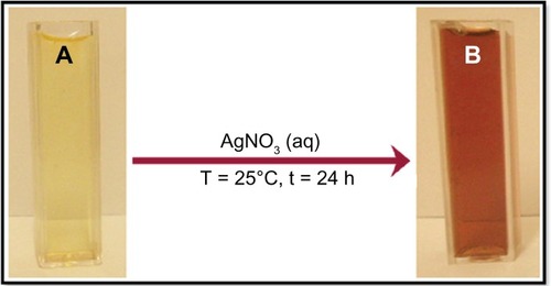
Figure 3 UV-visible absorption spectra of Curcuma longa (A) and silver (Ag)/C. longa emulsion (B) after 24 hours of stirring; Ag/C. longa emulsion (C) after 3 months.
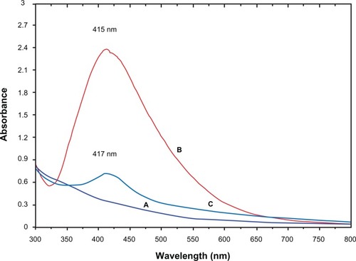
Figure 4 X-ray diffraction patterns of silver nanoparticles (Ag-NPs) synthesized in Curcuma longa for determination of Ag-NPs after 24 hours of stirring.
Abbreviation: XRD, X-ray diffration.
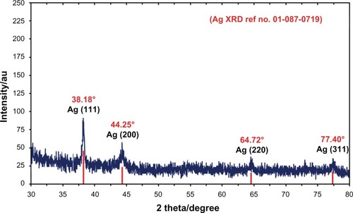
Figure 5 (A–D) Transmission electron microscopy images and corresponding size distribution of silver/Circuma longa after 24 hours of stirring.
Abbreviations: Std Dev, standard deviation; Ag-NPs, silver nanoparticles.
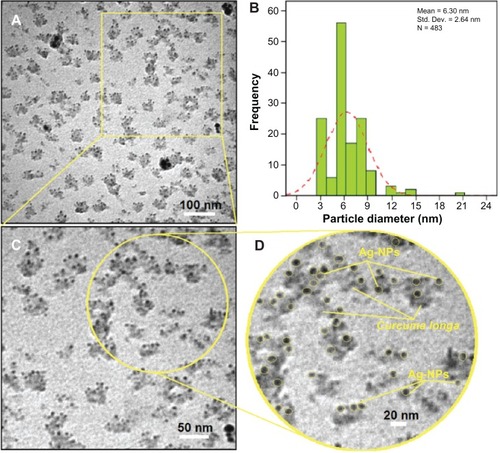
Figure 6 Scanning electron microscopy image and energy-dispersive X-ray fluorescence spectrometry spectra of Curcuma longa (A and B) and silver/C. longa (C and D) formation after 24 hours of stirring.
Abbreviation: Ag-NPs, silver nanoparticles.
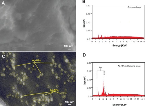
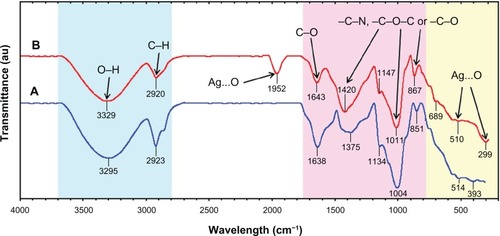
Figure 7 Fourier-transform infrared spectra for the Curcuma longa tuber-powder extract (A) and Ag/C. longa (B) after 24 hours from biosynthesis reaction.