Figures & data
Figure 1 SEM imaging of micro- and nanofiber electrospun PCL/PGLA tubular scaffolds designed for regenerating sciatic nerve transections. A) Tube lumen and B) zoomed details of the tube wall. Both nano- and microfibers are visible. Fiber links are obtained via partial solvent evaporation and polymer annealing subsequent to electrospinning in order to increase the overall prosthesis mechanical properties (image by courtesy of Joseph Lowery).
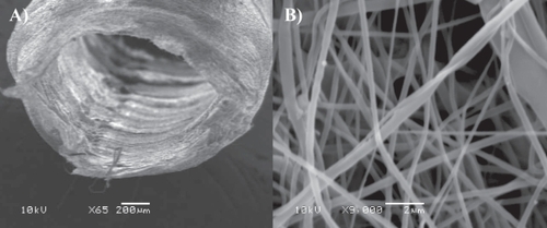
Figure 2 Human Neural Stem Cells cultured in a RADA16-I-BMHP1 3D scaffold (3 weeks in vitro). Cell nuclei are stained with DAPI (blue), neurons with βTubulin antibody (red), and astrocytes with GFAP antibody (green). In this long-term cultures neuronal morphologies resemble fairly mature neurons. A highly connected neuronal network is shown. Branched astrocytes also give evidence of differentiation of part of the stem cell progeny toward the astroglial phenotype.
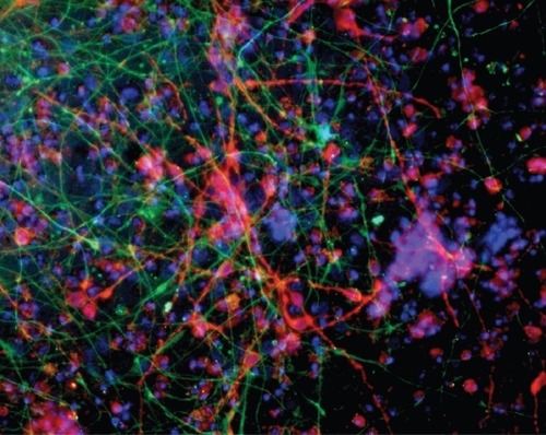
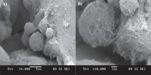
Figure 3 SEM imaging of a cluster of neural stem cells cultured in a RADA16-I-BMHP1 self-assembled scaffold. Low- (A) and high-magnification (B) images highlight cell bodies partially but tightly wrapped with functionalized nanofibers.