Figures & data
Table 1 Composition of experimental nanoparticles with their drug loading and entrapment efficiency
Figure 1 FTIR spectra of (A) stavudine; (B) PLGA; (C) PVA; (D) PVA and PLGA mixture; and (E) stavudine, PVA, and PLGA mixture.
Abbreviations: FTIR, Fourier transform infrared spectroscopy; PLGA, poly(d,l-lactic-co-glycolic acid); PVA, polyvinyl alcohol.
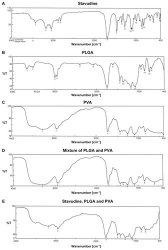
Table 2 Size distribution (Z-average [nm]), polydispersity index, zeta potential, and conductivity of experimental nanoparticulate formulations
Figure 2 FESEM photographs of formulations. (A) Formulation S1 prepared at 15,000 rpm shows that the particles were of micrometer sizes. (B) Formulation S2 prepared at 15,000 rpm shows that the particles were in micron range. (C) Formulation S3 prepared at 16,000 rpm shows that the particles were in nanometer range. (D) Formulation S4 prepared at 16,000 rpm shows that the particles were in nanometer range. (E) Formulation S5 prepared at 15,000 rpm shows that the particles were mostly in micrometer range. (F) SEM photograph of the gold nanoparticles shows that the particles were in submicron range with variable sizes.
Abbreviations: FESEM, field-emission scanning electron microscopy; SEM, scanning electron microscopy.
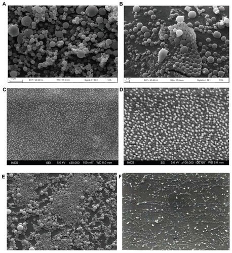
Figure 3 Size distribution of different formulations. (A) S1 (by intensity); (B) S2 (by intensity); (C) S3 (by intensity); (D) S4 (by intensity); (E) S5 (by intensity).
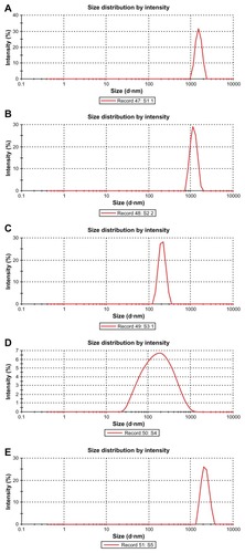
Figure 4 EDX data showing the presence of gold particles in gold-encapsulated polymeric nanoparticles.
Abbreviation: EDX, energy dispersive X-ray spectroscopy.
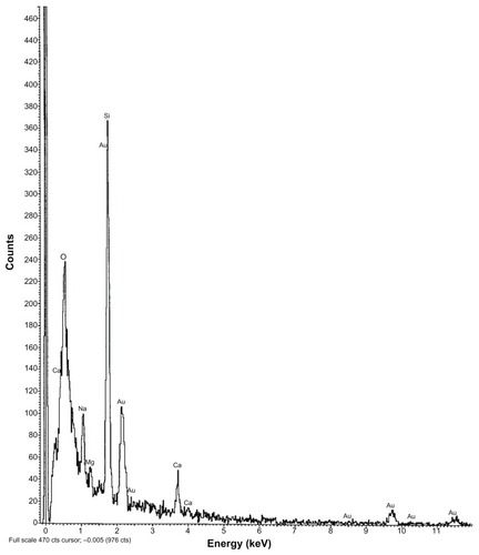
Figure 5 EDX data of colloidal gold nanoparticles alone.
Abbreviation: EDX, energy dispersive X-ray spectroscopy.
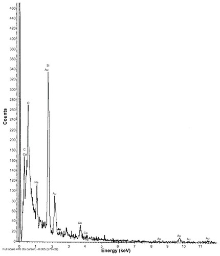
Figure 6 Release profile of didanosine from different formulations in phosphate-buffered saline (pH 7.4).
Note: Data show means (n = 3).
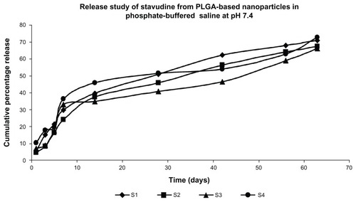
Table 3 Data of drug release kinetics of various formulations
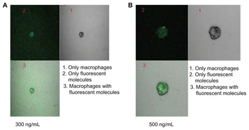
Table 4 Nanoparticle (S4) uptake by macrophages
Figure 7 Confocal microscopic image of macrophages treated with (A) stavudine nanoparticles (300 ng/mL of medium) for 4 hours, and (B) stavudine nanoparticles (500 ng/mL of medium) for 4 hours.