Figures & data
Figure 1 Chemical structure of compound 1 that forms the structure of ONTs and schematic illustrations of ONT (A) and Gd-chelated ONT (Gd-ONT) (B).
Note: Compound 1 consists of glycylglycine and myristic acid (molecular weight 342).
Abbreviations: Gd, gadolinium; ONTs, organic nanotubes.
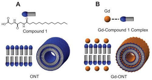
Figure 2 Field-emission scanning electron microscopy (FESEM) images of ONTs.
Abbreviation: ONTs, organic nanotubes.
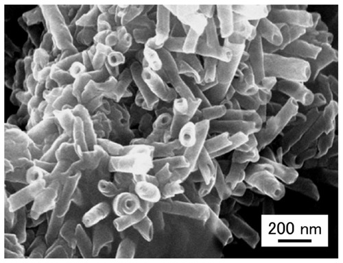
Figure 3 Effect of pH and ratio of CPT-11:ONT (w:w) on loading amount and loading efficiency of CPT-11 into ONTs at a CPT-11:ONT weight ratio of 0.25 (A) and at pH 4.0 (B) at room temperature.
Note: Each value represents mean ± SD (n = 3).
Abbreviations: ONTs, organic nanotubes; SD, standard deviation.
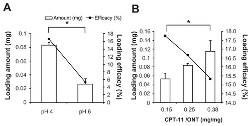
Figure 4 CPT-11 release from free CPT-11 and CPT-11/ONT in a dialysis tube in PBS at pH 5.5 and pH 7.4 at 37°C.
Note: Each value represents mean ± SD (n = 3).
Abbreviations: ONTs, organic nanotubes; PBS, phosphate-buffered saline; SD, standard deviation.
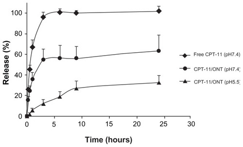
Figure 5 Biodistribution of free CPT-11 (4 mg/kg) and CPT-11/ONT (100 mg ONT/kg, equivalent to 2~3.5 mg CPT-11/kg) at 24 hours after a single intravenous injection into mice bearing C26 tumor. Tissue and tumor biodistribution of CPT-11 (A) and SN-38 (B).
Notes: Each value represents the mean ± SE (n = 3). *P < 0.05.
Abbreviations: ONTs, organic nanotubes; SE, standard error.
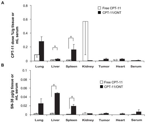
Figure 6 Biodistribution of Gd-ONT (50 mg ONT/kg, equivalent to 6.3 mg Gd/kg) at 3, 24, and 48 hours after a single intravenous injection into mice bearing C26 tumors.
Notes: Each value represents the mean ± SD (n = 3). *P < 0.05.
Abbreviations: ONTs, organic nanotubes; SD, standard deviation.
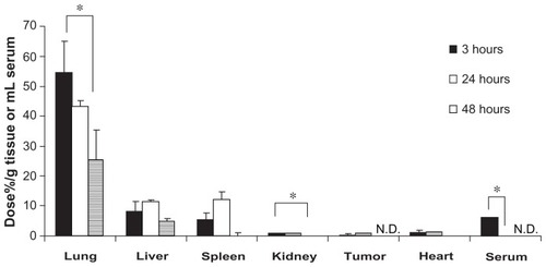
Figure 7 Biodistribution of MPs (25 mg/kg) at 3, 24 and 48 hours after a single intravenous injection into mice bearing C26 tumors.
Notes: Each value represents the mean ± SD (n = 3). *P < 0.05.
Abbreviations: MPs, microparticles; ONTs, organic nanotubes; SD, standard deviation.
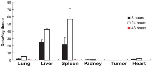
Figure 8 Biodistribution of DXR/ONT and MPs in lung tissues at 3 hours after a single intravenous injection into mice.
Notes: Fluorescence microscopy analyses confirming DXR/ONT, MPs and blood vessels (Hoechst 33342 staining) as red dots, green dots and blue fluorescence, respectively. Each saline was the control for red and green fluorescence. DXR/ONT was found outside of blood vessel in part, whereas MPs were found exclusively within the vessel lumen or associated with endothelial cells. Scale bars = 100 μm.
Abbreviations: DXR, doxorubicin; MPs, microparticles; ONTs, organic nanotubes.
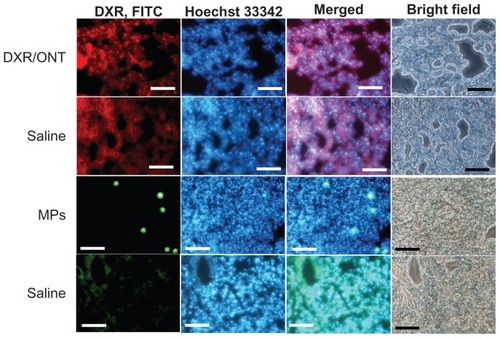
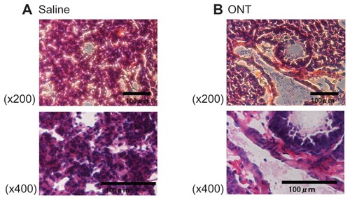
Figure 9 Hematoxylin and eosin-stained histological sections of lung tissue at 3 hours postinjection of saline (A) or ONTs (B).
Note: Scale bars = 100 μm.
Abbreviation: ONTs, organic nanotubes.