Figures & data
Figure 1 Differentially expressed miRNAs between exosomes derived from plasma of healthy people and patients with IS. (A) The extracellular vesicles were extracted from the plasma of patients with IS (n=3) or healthy people (n=3). Then, the extracellular vesicles were identified by TEM. (B) The expressions of CD9 and CD63 in extracellular vesicles from healthy people and patients with IS were detected by Western blot. (C) The differentially expressed miRNAs between the exosomes from healthy people and patients with IS were assessed using heatmap. (D) Volcano plots illustrating the differentially expressed miRNAs in IS. Red indicates a higher expression level, while green indicates a lower expression level.
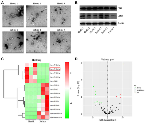
Figure 2 MiR-133a-3p upregulation reversed OGD/R-induced apoptosis in SH-SY5Y cells. (A) The expression of miR-133a-3p in plasma of healthy people or patients with IS was investigated by RT-qPCR. (B) SH-SY5Y cells were treated with OGD/R. The level of miR-133a-3p in SH-SY5Y cells was tested by RT-qPCR. (C) SH-SY5Y cells were transfected with NC or miR-133a-3p mimics. The expression of miR-133a-3p in SH-SY5Y cells was tested by RT-qPCR. (D) SH-SY5Y cells were treated with OGD/R, OGD/R + mimics-ctrl or OGD/R + miR-133a-3p mimics. The viability of SH-SY5Y cells was tested by CCK8 assay. (E) The LDH level in supernatants of SH-SY5Y cells was tested by ELISA. (F) The apoptosis in SH-SY5Y cells was tested by TUNEL staining. *P<0.05, **P< 0.01.
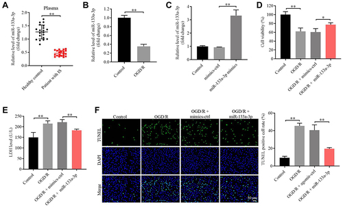
Figure 3 Exosomal miR-133a-3p derived from BMSCs reversed OGD/R-induced SH-SY5Y cell injury. SH-SY5Y cells were treated with OGD/R, OGD/R + BMSCs/NC-Exo or OGD/R + BMSCs/miR-133a-3p-Exo. (A) The level of miR-133a-3p in SH-SY5Y cells was investigated by RT-qPCR. (B) The viability of SH-SY5Y cells was tested by CCK8 assay. (C) The proliferation of SH-SY5Y cells was detected by EdU staining. (D) The LDH level in supernatants of SH-SY5Y cells was tested by ELISA. (E) The apoptosis in SH-SY5Y cells was tested by TUNEL staining. (F–H) The levels of MDA, SOD and GSH-Px in supernatants of SH-SY5Y cells were investigated by ELISA. **P< 0.01.
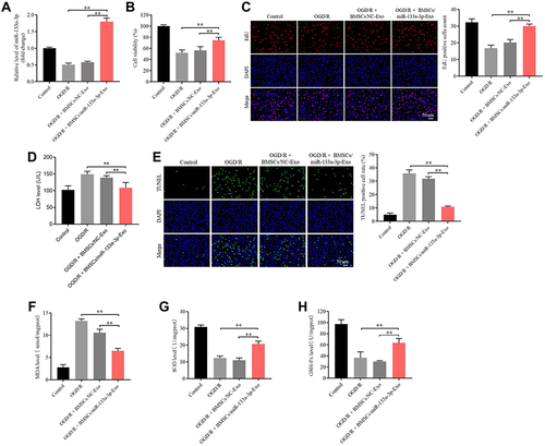
Figure 4 MiR-133a-3p directly targeted DAPK2. (A) Targetscan was used to predict the downstream mRNA of miR-133a-3p. (B) The relative luciferase activity in WT/MT-DAPK2 was detected by dual luciferase assay. (C) SH-SY5Y cells were transfected with mimics-ctrl or miR-133a-3p mimics. The levels of DAPK2 in SH-SY5Y cells were tested by RT-qPCR. (D) SH-SY5Y cells were treated with OGD/R, OGD/R + BMSCs/NC-Exo or OGD/R + BMSCs/miR-133a-3p-Exo. The protein levels of DAPK2, Akt, p-Akt, mTOR and p-mTOR in SH-SY5Y cells were investigated by Western blot. (E) The level of DAPK2 in plasma of patients with IS and healthy people was tested by RT-qPCR. (F) SH-SY5Y cells were treated with OGD/R, OGD/R + BMSCs/miR-133a-3p-Exo or OGD/R + BMSCs/miR-133a-3p-Exo + DAPK2 OE. The apoptosis of SH-SY5Y cells was tested by TUNEL staining. **P< 0.01.
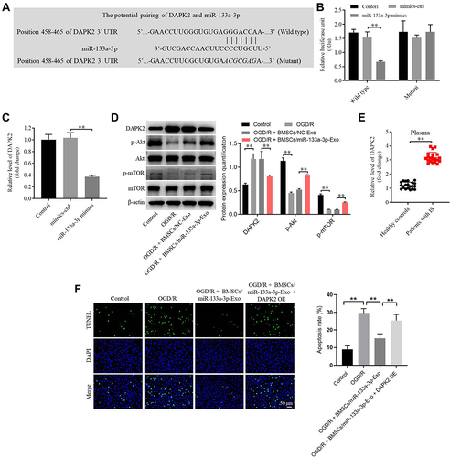
Figure 5 Exosomal miR-133a-3p derived from BMSCs alleviated OGD/R-induced SH-SY5Y cell apoptosis via inhibiting the autophagy. SH-SY5Y cells were treated with OGD/R, OGD/R + BMSCs/miR-133a-3p-Exo or OGD/R + BMSCs/miR-133a-3p-Exo + Rapamycin. (A) The protein level of Beclin-1 in SH-SY5Y cells was tested by Western blot. (B) The viability of SH-SY5Y cells was tested by CCK8 assay. (C) The apoptosis of SH-SY5Y cells was tested by TUNEL staining. **P< 0.01.
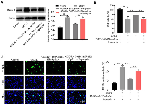
Figure 6 Exosomal miR-133a-3p derived from BMSCs markedly attenuated the symptom of CI/R injury in vivo. In vivo middle cerebral artery occlusion (MCAO) model in rats was established. At the end of the study, rats were sacrificed and the brain tissues were collected. Then, (A) the brain tissues were pictured, and TTC staining was performed to observe the severity of CI/R injury. (B) Cerebral infarction area was detected. (C) The expression of miR-133a-3p in tissues of rats was detected by RT-qPCR. (D) The protein levels of DAPK2, Akt, p-Akt, mTOR and p-mTOR in SH-SY5Y cells were investigated by Western blot. **P< 0.01.
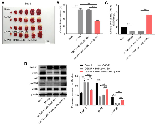
Figure 7 Exosomal miR-133a-3p derived from BMSCs attenuated MCAO-induced hippocampal neuronal degeneration. (A) H&E staining was performed to observe the histological changes in brain tissues of rats. (B) TUNEL staining was used to investigate the apoptosis in brain tissues of rats. (C) FJB staining was used to detect the hippocampal neuronal degeneration. **P< 0.01.
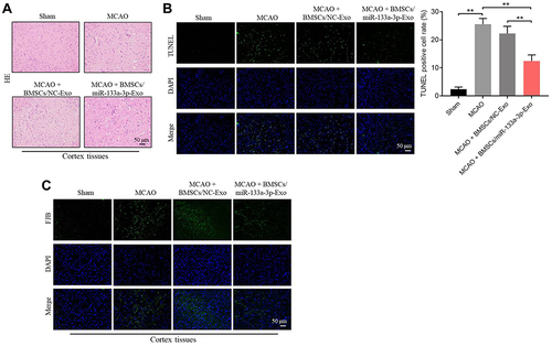
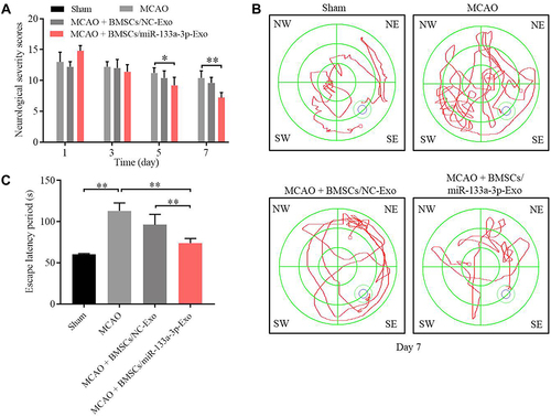
Figure 8 Exosomal miR-133a-3p derived from BMSCs significantly improved the memory capacity of MCAO rats. (A) The neurological severity scores of rats were tested. (B) Morris Water Maze assay was performed to test the memory capacity of rats. (C) The escape latency period of rats were evaluated. *P<0.05, **P< 0.01.