Figures & data
Figure 1 Functionalization of SPION-APTES, use and content of peptide and organic components. (A) Functionalization and separation procedure. (B) TGA of SPIONs, SPION-APTES and SPION-APTES-Pep and peptide alone. (C) Iron oxide and organic content ratio in % (w/w) for SPION-APTES-Pep (iron content was determined after evaporation by AES measurements). Experiments were performed in triplicates. Shown are the mean values with standard deviations.
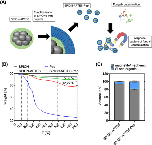
Figure 2 FTIR analysis of (A) SPION without coating, SPION-APTES and SPION-APTES-Pep. (B) Peptide (Pep).
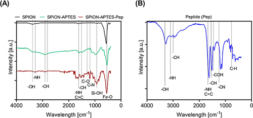
Figure 3 SPION-APTES-Pep after 12 weeks of storage. (A) Visual evaluation and UV-VIS spectra of SPION-APTES-Pep (100 µg Fe/mL). (B) FTIR measurements (kbr), (C) ζ-potential, (D) volume susceptibility and (E) Z-average of SPION-APTES-Pep freshly prepared and after 12 weeks of storage at 4 °C. Experiments were performed in triplicates. Shown are the mean results with standard deviation. Significance was calculated with a Mann–Whitney U-test. P values higher than 0.05 were considered non-significant (ns).
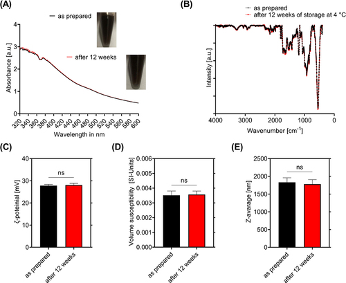
Figure 4 PIT-Analysis of cell cycle phases in (A) Jurkat T-cells, (B) THP-1 cells and (C) PBMCs after incubation with nanoparticles for 24 h. H2O served as negative control and DMSO 3% as positive control. Experiments were performed in three independent replications. Shown are the mean values with standard deviations. Significance was calculated with a one-way ANOVA test; ****p < 0.0001. P values higher than 0.05 were considered non-significant (ns). (D) shows a TEM-image of SPION-APTES-Pep engulfed by a phagocytic PBMC (red arrow) after 24 h of incubation with 400 µg Fe/mL.
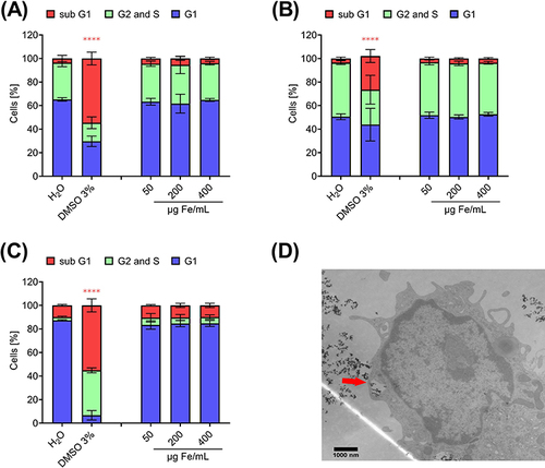
Figure 5 Removal of Candida albicans from aqueous (Ringer`s solution) samples. (A) Static system and (B) dynamic system with a flow rate of 10 mL/min using SPION-APTES-Pep. Different iron concentrations (300 µg Fe/mL and 100 µg Fe/mL) were tested. Untreated Candida albicans spiked samples served as positive control and medium without yeast as negative control. SPION-APTES-antibPep served as an additional control. Optical density was measured at 600 nm. Experiments were performed in triplicates of three independent repetitions. Shown are the mean values with standard deviation. Significance was calculated between treatment positive control and samples. Significances were calculated with a Kruskal–Wallis test; *p ≤ 0.05; ***p ≤ 0.001; ****p < 0.0001. P values higher than 0.05 were considered non-significant (ns). Simulation of the flow in the mix/separation camber (C and D) the magnetic flux density.
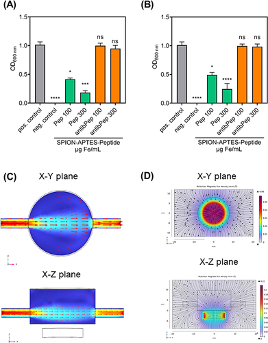
Figure 6 (A) Separation of Candida albicans (fluorescent strain) under static conditions in water-based media (Ringer`s solution). Before separation the cells are without any attachment at the cell wall. (B) After separation with SPION-APTES-Pep C. albicans cells from the magnetic pellet show attachment of particle clusters to the wall of the cells.
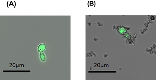
Figure 7 Removal and reculture of Candida albicans using SPION-APTES-Pep and SPION-APTES-antibPep. For the removal, the initial amount of Candida albicans in citrate-stabilized blood was set to a number of about 103 CFU/ mL. Experiments were performed in triplicates of three independent repetitions. Shown are the mean values with standard deviations. Significances were calculated between untreated samples with Candida albicans and treatment groups at the respective time. Significance was calculated with a Kruskal–Wallis test; *p ≤ 0.05; **p ≤ 0.001. P values higher than 0.05 were considered non-significant (ns).
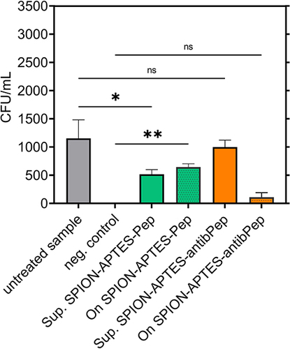
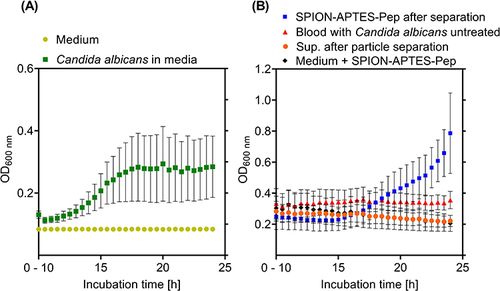
Figure 8 Growth and regrowth of Candida albicans. (A) Initial amount (before spiking into the culture medium) of Candida albicans was set to about 103 CFU/ mL. Samples were incubated for 24 h in culture medium. (B) Supernatant and SPION-APTES-Pep (in Ringer´s solution) after separation from citrate-stabilized blood were incubated in media (initial amount of candida was set to 103 CFU/ mL). Media with SPION-APTES-Pep and spiked blood samples are also shown. Samples were incubated for 24 h. Shown are the mean values from 10 h to 24 h of incubation with standard deviations.