Figures & data
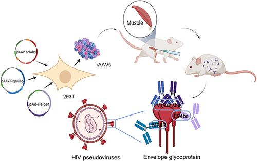
Figure 1 Antibody expression cassettes. (A) Schematic representation of bNAb constructs. Kpn I and Bgl II restriction sites are labeled atop the schematic. The expression vector components are labeled as follows: coding regions are located between the ITRs; CAG, promoters of the encoding protein; ss, human IL2 signal sequence leading to antibody secretion; HC, heavy chain antibody gene; LC, light chain antibody gene; Furin, furin cleavage site; 2A, FMDV 2A sequence. (B) Schematic representation of AAV expression plasmid.
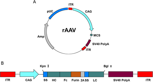
Figure 2 Characterization of purified rAAV8s. (A) Analysis of capsid proteins by SDS-PAGE indicating molecular weights of VP1 (∼87 kDa), VP2 (∼72 kDa), and VP3 (∼62 kDa). (B) The white arrows indicate the rAAV8 nano-vectors. Morphological analysis via TEM. Scale bar,100 nm.
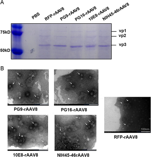
Figure 3 AAV-mediated antibody expression in HEK293T cells. RFP-rAAV8 was used as a negative control. The infection of 293T cells with RFP-rAAV8 was observed under a fluorescence microscope at 0h (A and D), 48h (B and E), and 72h (C and F). Scale, 50 μm. (G) Under non-reducing conditions, full-length dimerized antibodies and (H) under reducing conditions, the heavy chain of antibodies mediated by PG9-rAAV8, PG16-rAAV8, 10E8-rAAV8, and NIH45-46-rAAV8. (I) ID50 of expressed antibodies in HEK293T cell culture medium against tier 1 pseudoviruses.
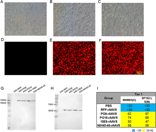
Figure 4 Gene expression mediated by rAAV8s in BALB/c mice. (A) Immunization schedule with a single injection of rAAV8 in BALB/c mice. (B) Biophotonic imaging of rAAV8 expressing RFP in BALB/c mouse muscle at 0, 4, 8, and 24 weeks. PBS is the corresponding control group. (C) The BG505 peptide was used to detect the concentration of PG9, PG16, and NIH45-46 antibodies. (D) Concentrations of the 10E8 antibody were assessed using gp41 protein.
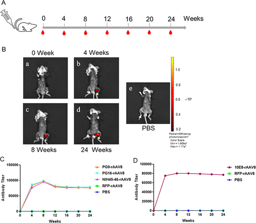
Figure 5 Evaluation of neutralizing activity of antibodies expressed by rAAV8s in BALB/c mice. Pre-immune sera and RFP-rAAV8 were used as negative controls. ID50 of expressed antibodies in BALB/c mice sera against tier 1 and tier 2 HIV-1 isolates.
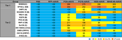
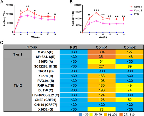
Figure 6 Antibody expression and neutralizing activity of antibodies expressed by combined rAAV8s. (A) The BG505 protein was used to detect the titer of co-expressed antibody. (B) The titer of the co-expressed antibody was assessed using the gp41 peptide. (C) ID50 of expressed antibodies in BALB/c mice sera against tier 1 and tier 2 HIV-1 isolates. *P < 0.05, **P < 0.01, ***P < 0.001.