Figures & data
Table 1 Commercialized NPs Have Been Explored as Contrast Agents for Molecular Imaging
Figure 1 Improving the imaging function by targeted nanoparticles. The common gastrointestinal tumor biomarkers overexpressed on cell membranes and the typical molecules/ligands used to modify the surface of nanoparticles for targeting imaging.
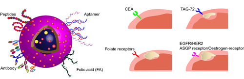
Table 2 NP Based Diagnostic Tests for GI Disorders
Figure 2 Nanoparticle combined with imaging technique for diagnostic of gastrointestinal disorder.Citation219
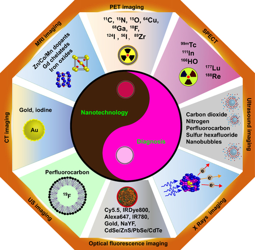
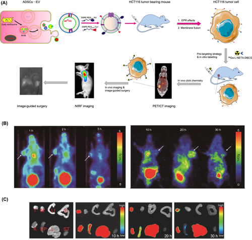
Figure 3 Multimodal PET and NIRF imaging based on ADSCs derived extracellular vesicles. (A) Schematic illustration showing the generation of exosome use for bio-imaging. (B) NIRF imaging of gastrointestinal tumor-bearing nude mice at different time points (1, 5, 10, 20, 30 and 50 h). The arrows indicate tumor sites. (C) In vitro tissue images at different time points (10, 20 and 30 h) after injection of Cy7-EV-N3.