Figures & data
Figure 1 Polymer composite scaffolds prepared with incorporation of micro/nanospheres or micro/nanofibers.
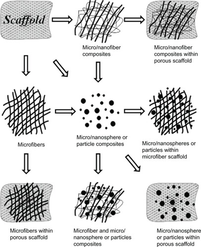
Figure 2 Incorporation of cells/DNA/growth factors into the scaffold matrix.
Abbreviation: DNA, deoxyribonucleic acid.
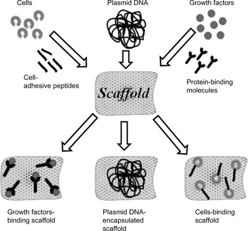
Table 1 Microcarrier fabrication methods using Fbg and Fbn and their biomedical applications
Figure 3 Microscopic images of Fbg microspheres.
Notes: (A) Light microscope image shows the microsphere sphericity and approximately uniform sizes, between 100–150 μm; (B) SEM image further confirmed sphericity; (C) Magnified image of (B), reveals the porous structures. Copyright © 2011, Springer Science+Business Media. Reproduced with permission from Rajangam T, Paik HJ, An SSA. Development of fibrinogen microspheres as a biodegradable carrier for tissue engineering. BioChip J. 2011;5(2):175–183.Citation9
Abbreviations: Fbg, fibrinogen; SEM, scanning electron microscope.
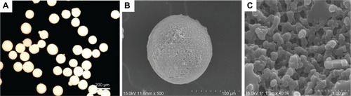
Figure 4 SEM images of Fbg nanospheres shown at different magnifications (image “A ×14,000” and image “B ×130,00” magnification).
Abbreviations: Fbg, fibrinogen; SEM, scanning electron microscope.
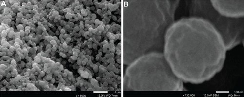
Figure 5 SEM images of Fbg microfibers fabricated from three different concentrations of Fbg.
Notes: Decreasing fiber diameter was observed with decreasing Fbg concentration. Images (A–C) show 200 μm, 150 μm, and 75 μm fibers, respectively, which were made from 15, 10, and 5 wt% Fbg, respectively. Images (D–F) represent highly aligned nanostructures, highly interporous fibers with aggregated structures, and less porous fibers with unaligned structure, respectively, and are the same as the lower magnification images in (A–C), respectively (inlet images scale bar is 1 μm). Copyright © 2012, Dove Medical Press Ltd. Reproduced with permission from Rajangam T, An SS. Improved fibronectin-immobilized fibrinogen microthreads for the attachment and proliferation of fibroblasts. Int J Nanomedicine. 2013;8:1037–1049.Citation51
Abbreviations: Fbg, fibrinogen; SEM, scanning electron microscope.
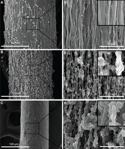
Figure 6 SEM images of an Fbg sheet reveal highly porous micro- and nanostructured networks (A and B shown at different magnifications).
Abbreviations: Fbg, fibrinogen; SEM, scanning electron microscope.
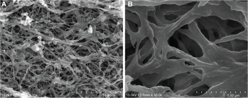
Figure 7 SEM images of a Fbg microtube. (A) The outer morphology of the tube shows the rough surfaces; (B) Magnified image of microtube shown in (A); (C) The tube’s inner surface was revealed to be much smoother than the outer surface; (D) The cross-sectional SEM image reveals the round shape, with approximately uniform wall thickness. (data unpublished).
Abbreviations: Fbg, fibrinogen; SEM, scanning electron microscope.
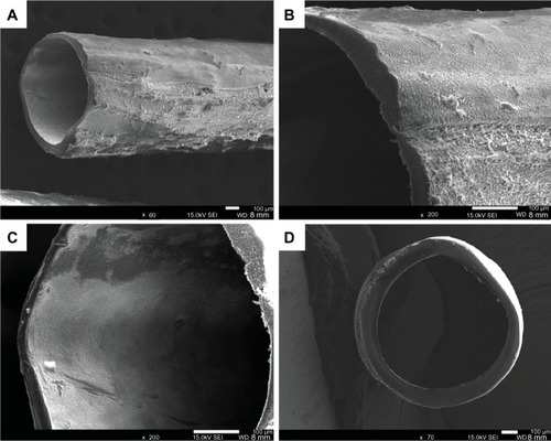
Table 2 Delivery systems for different biomolecules and cell types using Fbg/Fbn composites, and their tissue engineering applications
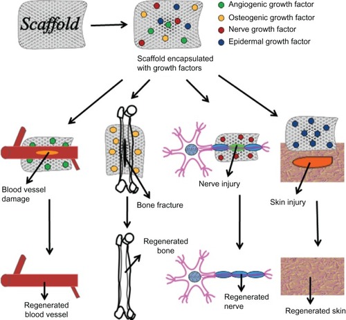
Figure 8 Specific growth factor–loaded scaffolds used for regeneration of various tissues.