Figures & data
Table 1 Preliminary phytochemical investigation of aqueous extract of C. zizanioides
Figure 1 Synthesis of silver and gold nanoparticles.
Notes: (A) AgNO3; (B) synthesized silver nanoparticles in brown color solution after 24 hours; (C) HAuCl4 solution; (D) synthesized gold nanoparticles in ruby red color after 24 hours.
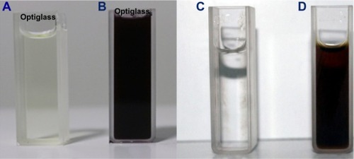
Figure 2 Time dependent absorption spectra of silver nanoparticles after the bioreduction of silver in the aqueous extract of Chrysopogon zizanioides.
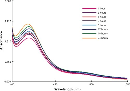
Figure 3 Time dependent absorption spectra of gold nanoparticles after the bioreduction with aqueous extract of Chrysopogon zizanioides.
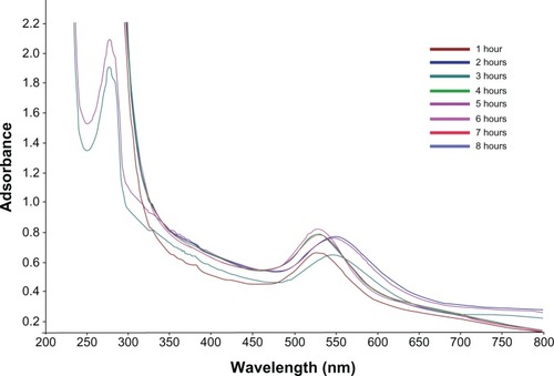
Figure 4 Scanning electron microscopy image of green silver nanoparticles synthesized by reduction of aqueous AgNO3 ions using Chrysopogon zizanioides extract.
Abbreviations: EHT, extra high tension; Mag, magnification; WD, working distance.
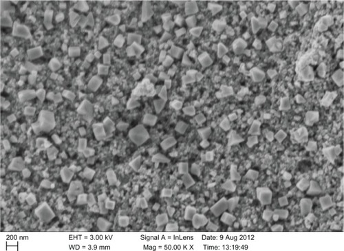
Figure 5 Scanning electron microscopy image of green gold nanoparticles synthesized by reducing aqueous AuCl4− ions using Chrysopogon zizanioides extract.
Abbreviations: Mag, magnification; WD, working distance; ETD, Everhart-Thornley detector; Vac, vacuum; HV, high voltage; Det, detector.
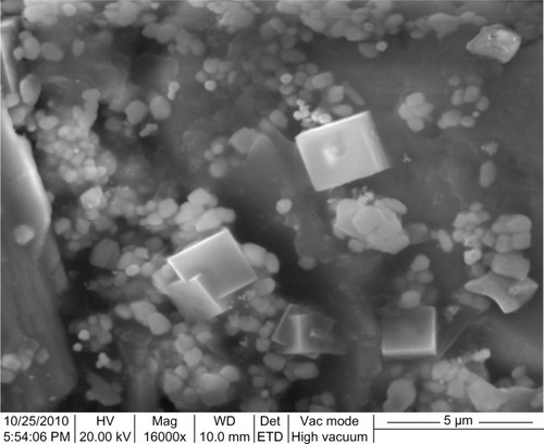
Figure 6 Energy dispersive X-ray spectrum of silver (Ag) nanoparticles.
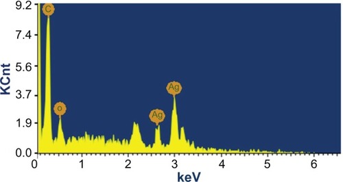
Figure 7 Energy dispersive X-ray spectrum of gold nanoparticles.
Abbreviation: Au, gold.
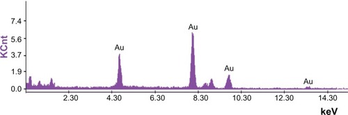
Figure 8 Fourier transform infrared spectroscopy spectrum of silver and gold nanoparticles.
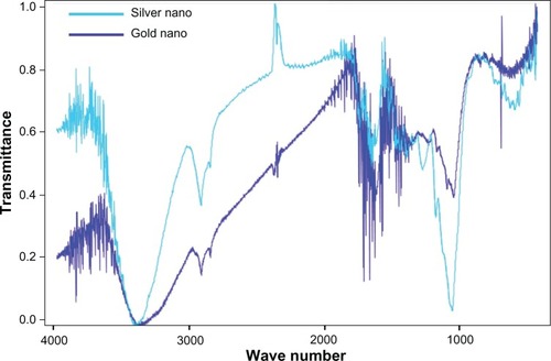
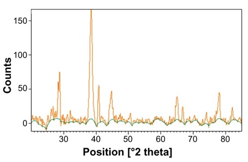
Figure 9 X-ray diffraction spectrum of green-synthesized silver nanoparticles.
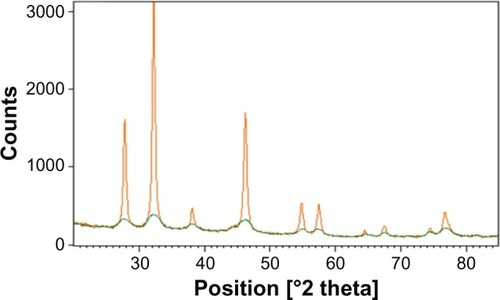
Figure 10 X-ray diffraction spectrum of green-synthesized gold nanoparticles.