Figures & data
Figure 1 Transmission electron micrographs of naked IONPs (A) and IONPs (arrows) in gel (B). Images of GBM-U87 cells in an untreated culture (C) and after 24 hours of incubation with IONPs 25 μg/mL (D).
Note: Arrowhead indicates intracellular localization of IONPs.
Abbreviation: IONPs, iron oxide nanoparticles.
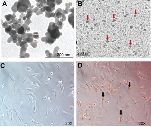
Figure 2 In vitro cell viability assay and cell counting with varying concentrations of IONPs (10, 25, 50, and 100 μg/mL) and incubation for 24, 48, and 72 hours (A). There was no significant difference in percent viability at any of the time points or concentrations in comparison with controls. The cytotoxicity induced by H2O2 10 mM was attenuated by 25 μg/mL IONPs (B). Live cell counting with the trypan blue exclusion method using different concentrations of IONPs (C), showing no significant difference in percentage of viable cells observed at any of the time points or concentrations in comparison with the control. Cytotoxicity induced by H2O2 10 mM was attenuated by 25 μg/mL IONPs (D).
Notes: Statistical significance (P < 0.05) is shown by aControl versus IONP, H2O2, IONP + H2O2; bH2O2 versus NP + H2O2.
Abbreviations: IONPs, iron oxide nanoparticles; H2O2, hydrogen peroxide.
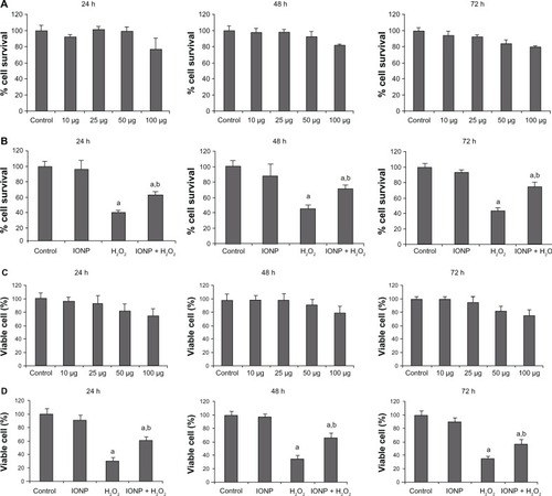
Table 1 Effect of iron oxide nanoparticle implantation and/or exposure to a magnetic field on general body condition
Figure 3 Effect of IONP implantation and MF exposure on mean (± SD) BBB score (A) and mean (± SD) time of stay on the moving Rotarod (B). Significant recovery was observed in both these tests after IONP implantation and MF exposure starting from week 1 which continued throughout the observation period. Mean (± SD) hind paw withdrawal latency (C) and urinary bladder function (D) were also significantly better in the NP + MF group.
Notes: Statistical significance (P < 0.05) is shown by aSCI versus MF, NP, NP + MF groups; bNP versus MF/NP + MF groups; cMF versus NP + MF group.
Abbreviations: BBB, Basso, Beattie, and Bresnahan locomotor rating; IONPs, iron oxide nanoparticles; SD, standard deviation; SCI, spinal cord injury group; MF, SCI + magnetic field group; NP, SCI + IONP implantation group; NP + MF, SCI + IONP implantation + magnetic field group.
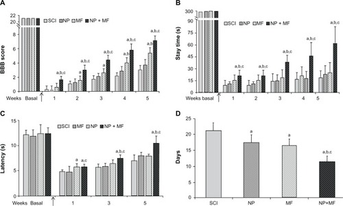
Figure 4 Histological analysis of lesion site. Representative images of stained sections from each group showing extent of the lesion (A) and presence of viable motor neurons (arrowhead) (B) in the NP + MF group. Masson’s trichrome staining for collagenous connective tissue (C). Dense collagenous staining was observed in the SCI and NP groups (D), indicating scar formation; this was significantly less in the MF group and was absent in the NP + MF group.
Notes: Scale bar (A and C) = 1000 μm and (B and D) = 100 μm.
Abbreviations: IONPs, iron oxide nanoparticles; SCI, spinal cord injury group; MF, SCI + magnetic field group; NP, SCI + IONP implantation group; NP + MF, SCI + IONP implantation + magnetic field group.
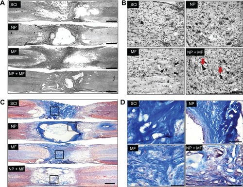
Figure 5 Mean (± SD) total tissue damage volume was significantly less in all groups in comparison with the SCI group, but least in the NP + MF group.
Notes: Statistical significance (P < 0.05) is shown by aSCI versus MF, NP, and NP + MF groups; bNP versus MF and NP + MF groups; cMF versus NP + MF group.
Abbreviations: IONPs, iron oxide nanoparticles; SCI, spinal cord injury group; SD, standard deviation; MF, SCI + magnetic field group; NP, SCI + IONP implantation group; NP + MF, SCI + IONP implantation + magnetic field group.
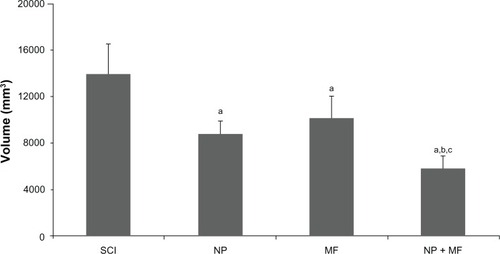
Figure 6 Prussian blue staining for iron (A), showing that IONPs diffused out of the gel and concentrated in tissues near the site of the lesion. This aggregation of IONPs was high in the presence of MF in the NP + MF group. The arrowhead indicates the location of IONPs inside the motor neurons and axons (B). Transmission electron microscope images of tissues showed cytoplasmic localization of IONPs (arrows) (C).
Notes: Scale bar (A) = 100 μm, (B) = 10 μm, and (C) = 500 nm.
Abbreviations: IONPs, iron oxide nanoparticles; SCI, spinal cord injury group; MF, SCI + magnetic field group; NP, SCI + IONP implantation group; NP + MF, SCI + IONP implantation + magnetic field group.
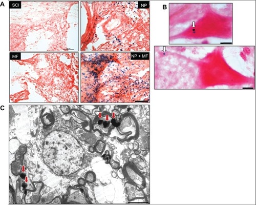
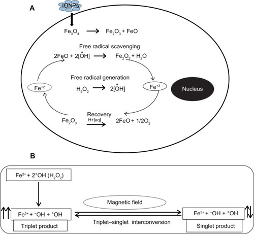
Figure 7 Proposed mechanism for free radical scavenging by IONPs (A) and interaction of radical pairs with MF (B).
Notes: The presence of mixed valence states for Fe2+ and Fe3+ in the Fe3O4 acts as an antioxidant, enabling the IONPs to scavenge free radicals from the cell. Hydroxyl radical (°OH) produced from H2O2 reacts with an electron donor (Fe2+) and results in generation of a pair of radicals (Fe3+ + −OH + °OH) by electron transfer from Fe2+ to °OH. An external MF affects interconversion between singlet and triplet states of the radical pair. We believe that low frequency MF leads to spin relaxation and favors generation of a singlet product and prevents generation of oxidative stress by interaction of the triplet product with biomolecules in the cell. Another set of chemical reactions between the ions in the cell and Fe3O4 then appear to be involved in reversing the oxidation state from Fe3+ to Fe2+. We believe that this is indicative of a cyclical regenerative or autocatalytic reaction on the part of IONPs.
Abbreviations: IONPs, iron oxide nanoparticles; Fe2+, ferrous ion; Fe3+, ferric ion; MF, magnetic field; H2O2, hydrogen peroxide; −OH, hydroxyl ion; °OH, hydroxyl radical; Fe3O4, iron (II,III) oxide.