Figures & data
Figure 1 Preparation scheme for doxorubicin-loaded MPEG-PCL nanoparticles. There were two steps involved in the preparation of Cur-Dox/MPEG-PCL. First, Cur/MPEG-PCL was prepared by a self-assembly method. Second, Cur-Dox/MPEG-PCL micelles were prepared by pH-induced self-assembly method.
Abbreviations: Cur, curcumin; Dox, doxorubicin; MPEG, methoxy poly(ethylene glycol); PCL, poly(caprolactone).
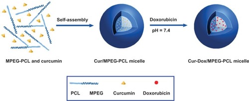
Figure 2 Characterization of Cur-Dox/MPEG-PCL micelles showing their particle size. The Cur-Dox/MPEG-PCL micelles had a very narrow particle size distribution (polydispersity index 0.12) with a mean particle size of 38.4 ± 1.2 nm, determined by dynamic light scattering (A). Cur-Dox/MPEG-PCL micelles with a zeta potential of −0.269 mV (B). Transmission electronic microscopic images of Cur-Dox/MPEG-PCL micelles showing a spherical morphology with a mean diameter of about 27 nm (C). Appearance of Dox/MPEG-PCL (D) and Cur/MPEG-PCL (E) micelles in aqueous solution. Appearance of Cur-Dox/MPEG-PCL, Dox/MPEG-PCL, and Cur/MPEG-PCL diluted in water (F). Cur-Dox/MPEG-PCL, Dox/MPEG-PCL, Cur/MPEG-PCL and water are shown from left to right. Data are shown as the mean ± standard.
Abbreviations: Cur, curcumin; Dox, doxorubicin; MPEG, methoxy poly(ethylene glycol); PCL, poly(caprolactone); NS, normal saline; M, MPEG-PCL; C, Cur/MPEG-PCL; D, Dox/MPEG-PCL; C+D: Cur-Dox/MPEG-PCL.
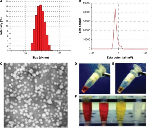
Figure 3 Release profile of curcumin or doxorubicin micelles in vitro studied using a dialysis method. Doxorubicin was released from Dox/MPEG-PCL (A), curcumin was released from Cur/MPEG-PCL (B), doxorubicin was released from Cur-Dox/MPEG-PCL (C) and curcumin was released from Cur-Dox/MPEG-PCL (D) over an extended period. Data are shown as the mean ± standard error of the mean.
Abbreviations: Cur, curcumin; Dox, doxorubicin; MPEG, methoxy poly(ethylene glycol); PCL, poly(caprolactone).
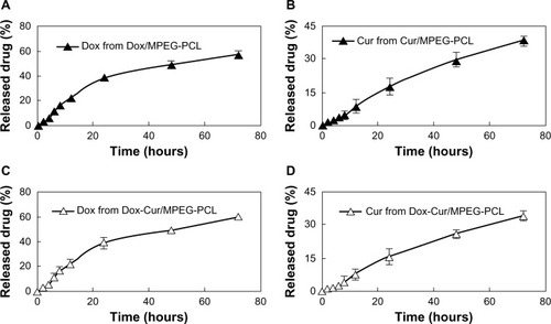
Figure 4 Synergistic antitumor effect of curcumin and doxorubicin on an LL/2 tumor cell line. The MTT assay (A), showed that the combination group (ratio of curcumin to doxorubicin, 1:1) was more effective than curcumin and doxorubicin alone at concentrations of 0.75 μg/mL, 1.5 μg/mL, and 3 μg/mL at 24 hours. Flow cytometry determination of cell apoptosis (B) showed that the curcumin-induced apoptosis efficiency was 9.34%, doxorubicin-induced apoptosis was 10.55%, and apoptosis induced by combination therapy was 16.08% at a concentration of 0.75 μg/mL; the 1.5 μg/mL concentration of curcumin induced an apoptosis efficiency of 9.57%, the doxorubicin-induced apoptosis was 11.33%, and combination therapy induced apoptosis of 20.02%; at the 3 μg/mL concentration, curcumin-induced apoptosis efficiency was 11.54%, doxorubicin-induced apoptosis was 15.53%, and apoptosis induced by combination therapy was 29.13%. Data are shown as the mean ± standard error of the mean. *P < 0.05 between groups.
Abbreviations: Cur, curcumin; Dox, doxorubicin; MPEG, methoxy poly(ethylene glycol); PCL, poly(caprolactone); groups are as follow: NS, normal saline treated group; M, MPEG-PCL micelles treated group; C, Cur/MPEG-PCL micelles treated group; D, Dox/MPEG-PCL micelles treated group; C+D: Cur-Dox/MPEG-PCL micelles treated group.
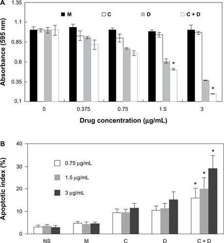
Figure 5 Antiangiogenesis effect of Cur-Dox/MPEG-PCL. Normal embryos (A), embryos treated with empty MPEG-PCL (B), embryos treated with Cur/MPEG-PCL micelles (C), embryos treated with Dox/MPEG-PCL micelles (D), and embryos treated with Cur-Dox/MPEG-PCL micelles (E). Embryos treated with Cur-Dox/MPEG-PCL micelles showing defective vascular formation of variable severity, with intersegmental vessels either sprouting abnormally or failing to form in comparison with control embryos.
Abbreviations: Cur, curcumin; Dox, doxorubicin; MPEG, methoxy poly(ethylene glycol); PCL, poly(caprolactone).
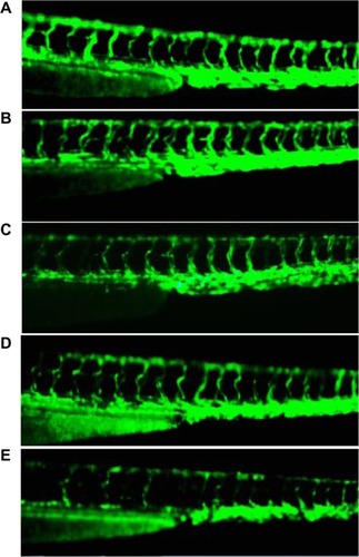
Figure 6 Anticancer activity in vivo. A C57BL/6J mouse tumor model was established by subcutaneous injection with LL/2 cells. Mice were treated with normal saline (NS), MPEG-PCL (M) micelles, Cur/MPEG-PCL (Cur) micelles (curcumin 5 mg/kg), Dox/MPEG-PCL (Dox) micelles (doxorubicin 5 mg/kg), Cur-Dox/MPEG-PCL (C + D) micelles (curcumin and doxorubicin 5 mg/kg), respectively. A statistically significant difference was found in tumor volume (*P < 0.05) between the Cur-Dox/MPEG-PCL group and other treatments alone (A); the same result was found for tumor weights (B). Points, mean (n = 8); bars, standard deviation.
Abbreviations: Cur, curcumin; Dox, doxorubicin; MPEG, methoxy poly(ethylene glycol); PCL, poly(caprolactone).
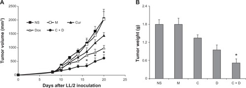
Figure 7 Apoptosis of lung cancer cells was detected using TUNEL analysis. The percentage of apoptosis was determined by counting the number of apoptotic cells and dividing by the total number of cells in the field (five high power fields per slide). Normal saline group (A), MPEG-PCL group (B), Cur/MPEG-PCL group (C), Dox/MPEG-PCL group (D), Cur-Dox/MPEG-PCL group (E), and Percent apoptosis in each group (F). Treatment with Cur-Dox/MPEG-PCL resulted in significantly increased apoptosis compared with that of other groups. *P, 0.05. Bars indicate standard deviation; columns show the mean.
Abbreviations: Cur, curcumin; Dox, doxorubicin; MPEG, methoxy poly(ethylene glycol); PCL, poly(caprolactone); NS, normal saline; M, MPEG-PCL; C, Cur/MPEG-PCL; D, Dox/MPEG-PCL; C+D: Cur-Dox/MPEG-PCL.
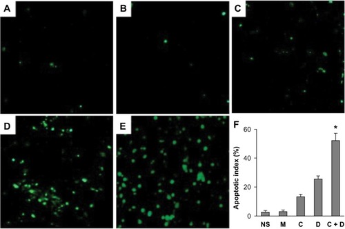
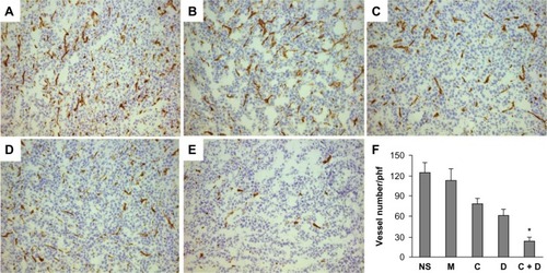
Figure 8 Normal saline group (A), MPEG-PCL group (B), Cur/MPEG-PCL group (C), Dox/MPEG-PCL group (D), Cur-Dox/MPEG-PCL group (E), and vessel density of tumor tissues in each group (F). *P<0.05. Bars indicate standard deviation; columns show the mean.
Abbreviations: Cur, curcumin; Dox, doxorubicin; MPEG, methoxy poly(ethylene glycol); PCL, poly(caprolactone); phf, per high field; NS, normal saline; M, MPEG-PCL; C, Cur/MPEG-PCL; D, Dox/MPEG-PCL; C+D: Cur-Dox/MPEG-PCL.