
Figures & data
Figure 1 Macroscopic and microscopic changes of the IVDD during degeneration. Characteristic changes of the NP’s ECM after degeneration are illustrated in the dashed circles. ECM of degenerated discs (dashed red circle) shows shorter aggrecan macromolecules and more collagen type I fibres (thicker fibre bundles) than collagen type II (thinner collagen bundles), which are largely abundant in the ECM of healthy discs (dashed green circle). Reprinted from Costăchescu B, Niculescu AG, Teleanu RI, et al. Recent advances in managing spinal intervertebral discs degeneration. Int J Mol Sci. 2022;23(12):6460. Creative Commons.Citation78
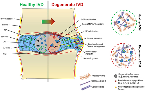
Figure 2 A new injectable thermosensitive hydrogel with ROS-sensitive MR409-loaded vesicles demonstrates sustained controlled release and biocompatibility. (A) Schematic illustration of the thermosensitive hydrogel loaded with ROS-responsive PPS-PEG vesicles for controlled release of MR409. (B) Hydrodynamic diameters and (C) MR409-loading efficiencies of PPS-PEG vesicles. (D) Viscosity of the PLGA-PEG-PLGA solution containing MR409-loaded vesicles as a function of temperature. (E) G’ and G” of the PLGA-PEG-PLGA solution containing MR409-loaded vesicles as a function of temperature. (F) Change in the morphology of MR409-loaded vesicles in the presence of external H2O2 (100 µM). (G) Cumulative release of MR409 from PPS-PEG vesicles or hydrogel containing vesicles in the presence of 100 µM H2O2. (H and I) Images of live/dead cell staining (H) and quantitation (I) of rat NP cells cultured with hydrogel confirming good biocompatibility. Reprinted from Zheng Q, Shen H, Tong Z, et al. A thermosensitive, reactive oxygen species-responsive, MR409-encapsulated hydrogel ameliorates disc degeneration in rats by inhibiting the secretory autophagy pathway. Theranostics. 2021;11(1):147–163. Creative Commons.Citation85
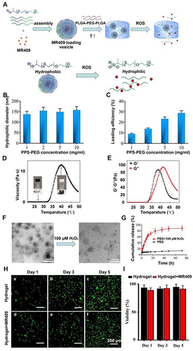
Figure 3 (A) Schematic illustration for formation of miRNA/PGPC polyplex micelles. (B) Encapsulation of miRNA/PGPC polyplexes in PEG hydrogels in an injectable manner and molecular mechanism of MMP-2 silence in nucleus pulposus cells for fibrosis inhibition. (C) Injection sites in the IVDs of rabbits. Reprinted from Feng G, Zha Z, Huang Y, et al. Sustained and Bioresponsive Two-Stage Delivery of Therapeutic miRNA via Polyplex Micelle-Loaded Injectable Hydrogels for Inhibition of Intervertebral Disc Fibrosis. Adv Healthc Mater. 2018 Nov;7(21):e1800623. VCH Verlag GmbH & Co. KGaA, Weinheim.Citation99
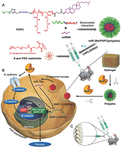
Figure 4 General schematic of synthesis of esterase-responsive ibuprofen nano-micelles (PEG-PIB) to pre-modify embryo-derived long-term expandable nucleus pulposus progenitor cells (NPPCs) for synergistic transplantation in intervertebral disc degeneration. Reprinted from Xia KS, Li DD, Wang CG, et al. An esterase-responsive ibuprofen nano-micelle pre-modified embryo derived nucleus pulposus progenitor cells promote the regeneration of intervertebral disc degeneration. Bioact Mater. 2023;21:69–85. Creative Commons.Citation102
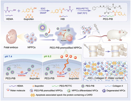
Figure 5 In vivo therapeutic effects in a rat IVDD model. (A) Schematic illustration showing the overall processes of animal experiments. (B) X-ray and (C) MRI images of rat coccygeal vertebral discs at different time points indicated that the height and water content of the degenerative intervertebral disc were significantly improved after treatment with the Hydrogel/TA NPs@ant. (D and E) The changes in DHI and (F) MRI grade at 4 and 8 w quantitatively confirmed the repair effect of IVDD implanted with Hydrogel/TA NPs@ant. (G) Representative images of H&E, safranin-O/fast green, Col II, MMP-13 and TNF-α immunohistochemical staining of different treatments at 4 and 8 w (scale bars, 1 mm (for H&E, safranin-O/fast green), 50 μm (for immunohistochemical staining)). The histological results further confirmed the role of the Hydrogel/TA NPs@ant in improving the metabolic balance of the ECM in degenerative nucleus pulposus and inhibiting the infiltration of inflammatory factors. (H) The histological grades in different groups further proved the synergistic effect of antagomir-21, TA NPs and the hydrogel on nucleus pulposus regeneration. (I) Heatmap represents the total intensity of Col II, MMP-13 and TNF-α immunohistochemical staining in nucleus pulposus of different treatments at 8 w. Reprinted from Biomaterials, volume: 298, Wang Y, Wu Y, Zhang B, et al. Repair of degenerative nucleus pulposus by polyphenol nanosphere-encapsulated hydrogel gene delivery system. 122132. Copyright 2023, with permission from Elsevier.Citation113
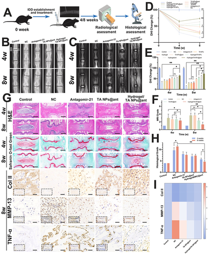
Figure 6 Various properties of the temperature-sensitive material hyaluronan-poly(N-isopropylacrylamide) HA-pNIPAM. Reprinted with permission from Mortisen D, Peroglio M, Alini M, Eglin D. Tailoring thermoreversible hyaluronan hydrogels by “click” chemistry and RAFT polymerization for cell and drug therapy. Biomacromolecules. 2010;11(5):1261–1272. Copyright {2010} American Chemical Society.Citation114
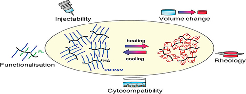
Figure 7 Schematic illustration of miRNA-based gene therapy delivered by a thermo-responsive vector for intervertebral disc degeneration (IVDD). (A) The chemical structure of cationic block copolymers: PNIPAM-b-PAsp (DET) and PEG-b-PAsp (DET). (B) The mixed cationic block copolymers (MCBC) PNIPAM-b-PAsp (DET) and PEG-b-PAsp (DET) as vectors for miRNA to form mixed polyplex micelles. With a temperature increase from room temperature (25 °C) to physiological temperature (37 °C), thermo-sensitivity of PNIPAM segments become hydrophobic and form more compact mixed polyplex micelles with hydrophobic and hydrophilic heterogeneous surfaces. The mixed cationic block copolymers delivered miRNA-25-3p for IVDD gene therapy. Reprinted from Huang Y, Huang L, Li L, et al. MicroRNA-25-3p therapy for intervertebral disc degeneration by targeting the IL-1β/ZIP8/MTF1 signaling pathway with a novel thermo-responsive vector. Ann Transl Med. 2020;8(22):1500. Creative Commons.Citation125
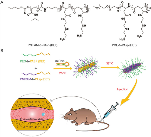
Figure 8 Schematic showing the process of photo-curing of hMSC encapsulated within p(HEMA-co-APMA) g PAA hydrogels: (A) procedure of Three-dimensional hMSC encapsulation in polymer to produce hydrogel; (B) representative image of a 13.5% (w/v) photo-cured hydrogel with dimensions 1 cm Ø and 3 mm thick). Reprinted from Kumar D, Gerges I, Tamplenizza M, Lenardi C, Forsyth NR, Liu Y. Three-dimensional hypoxic culture of human mesenchymal stem cells encapsulated in a photocurable, biodegradable polymer hydrogel: a potential injectable cellular product for nucleus pulposus regeneration. Acta Biomater. 2014;10(8):3463–3474. Creative Commons.Citation139
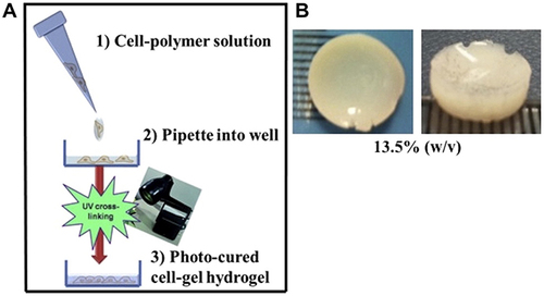
Figure 9 A schematic illustration for the experiment design and the fabrication process of RES-loaded, CDH2-decorated nanobubbles. Reprinted from Shen J, Zhuo N, Xu S et al. Resveratrol delivery by ultrasound-mediated nanobubbles targeting nucleus pulposus cells. Nanomedicine (Lond). 2018 Jun;13(12):1433–1446.Citation154 Copyright 2018 Future Medicine Ltd; permission conveyed through Copyright Clearance Center, Inc.
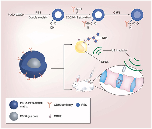
Figure 10 Schemes depicting the different types of magnetic responsive materials obtained from the doping of various polymers with magnetic particles and illustration of their response when exposed to a static magnetic field (H dc) or to an alternating magnetic field (H ac). From left to right: composites made from elastomeric polymers can be deformed in homogeneous fields or gradients in a controlled fashion (A-C); MRPC particles made of polymers designed for biomedical applications can be used for magnetic guidance for drug delivery or separation purpose (D–F); MRPC from thermo-responsive polymers can be activated by magnetic induction using alternating fields (G and H). Reprinted from Thévenot J, Oliveira H, Sandre O, Lecommandoux S. Magnetic responsive polymer composite materials. Chem Soc Rev. 2013 Sep 7;42(17):7099–116.Citation159 Copyright 2013 Royal Society of Chemistry; permission conveyed through Copyright Clearance Center, Inc.
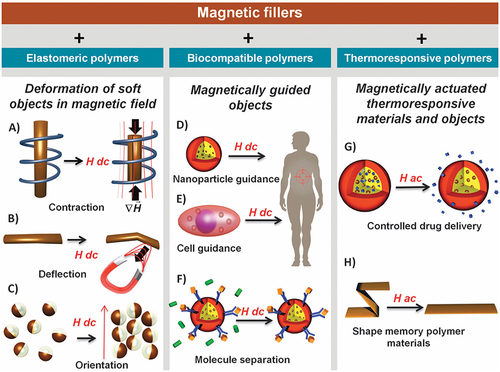
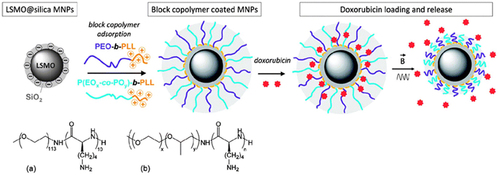
Figure 11 Preparation of magnetic responsive drug delivery systems (DDS) based on lanthanum strontium manganese oxide (LSMO) MNPs and polymer brush with a tunable LCST (top left). Chemical structures of the block copolymers used: (a) poly (ethylene glycol)-b-poly(lysine), and (b) poly (ethylene glycol-co-propylene glycol)-b-poly(lysine), with x = 6 and y = 29. Principle of drug loading and alternating magnetic field (AMF) triggered drug release (right). Reprinted from Louguet S, Rousseau B, Epherre R, et al. Thermoresponsive polymer brush-functionalized magnetic manganite nanoparticles for remotely triggered drug release. 10.1039/C2PY20089A. Polym Chem. 2012;3(6):1408–1417.Citation165 Copyright 2013 Royal Society of Chemistry; permission conveyed through Copyright Clearance Center, Inc.
Data Sharing Statement
The datasets used and/or analyzed during the current study are available from the corresponding author on reasonable request.