Figures & data
Figure 1 Biogenesis, markers, and contents of exosomes. The canonical biogenesis pathway of exosomes involves the invagination of the plasma membrane, with the ESCRT facilitating cargo loading to form MVBs. Subsequent fusion of MVBs with the cellular membrane releases exosomes. Biomarkers for exosomes mainly include CD9, CD63, CD81, Alix, HSP70, and TSG101. The cargoes of exosomes are comprised of DNA, RNA (mRNA, miRNA, circRNA, and IncRNA), proteins, and lipids. ESCRT, endosomal sorting complex for transport; MVBs, multivesicular bodies; HSP70, heat shock protein 70; mRNAs, messenger RNAs; miRNAs, microRNA; circRNAs, circular RNAs; IncRNAs, long non-coding RNAs. (Figure created using Figdraw).
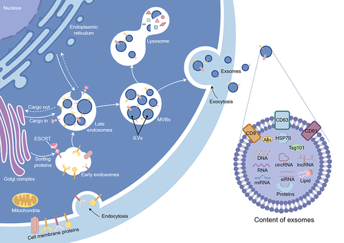
Figure 2 Four stages of normal wound healing. The normal wound healing process consists of four distinct overlapping stages, namely hemostasis and coagulation, the inflammatory stage, the proliferative stage, and remodeling. (Figure created using Figdraw).
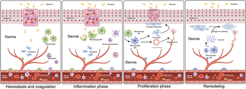
Figure 3 Various factors for diabetic wounds exhibit delayed healing. Multiple factors collectively contribute to the prolonged non-healing of diabetic wounds, mainly including the cytotoxicity of HG, microvascular complications, neuropathy, disruption of antimicrobial barriers, inflammation, and immune deficiency. HG, high glucose. (Figure created using Figdraw).
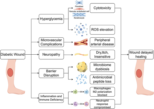
Table 1 Summary of ADSC-Exos Promoting Diabetic Wound Healing in vivo
Figure 4 Promotion effect of ADSC-exos on skin cell functions. Existing cell experimental results indicate that under high-glucose conditions, ADSC-exos impacted various biological functions of skin cells, including proliferation, migration, angiogenesis, and anti-apoptosis, and could promote macrophage M2 polarization. Ultimately, ADSC-exos promote the diabetic wound healing. Exosomes from hypoxia, overexpression of specific circRNAs, or NFIC transfection pretreated ADSCs also present robust capabilities of healing-promoting effect. (Figure created using Figdraw).
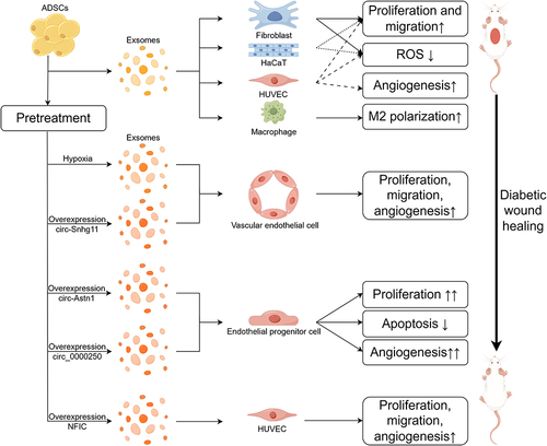
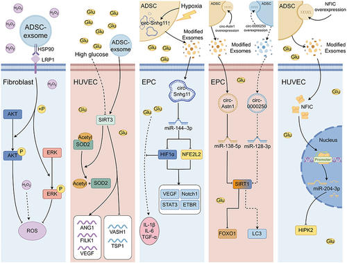
Figure 5 Partial molecular mechanisms underlying the pro-healing effects of ADSC-exos in different cells. The ADSC-exo membrane protein HSP90 is bound to the fibroblast membrane protein LPR1 and promote AKT/ERK phosphorylation. ADSC-exos promote SIRT3 expression, downregulate the acetylation level of SOD2, and simultaneously upregulate ANG1, FILK1, and VEGF, while downregulate VASH1 and TSP1 in HUVECs. The ADSC-exo circ-Snhg11 is promoted by hypoxia. And circ-Snhg11 inhibit miR-144-3p, thereby promoting the protein expression of HIF-1α and NFE2L2, which in turn promote the expression of downstream proteins such as VEGF, Notch1, STAT3 and ETBR in EPCs. HIF-1α inhibits HG-induced inflammatory factors. The molecular mechanism of circRNA overexpression modified ADSC-exos in EPCs is as follows: circ-Astn1 inhibits miR-138-5p, relieves the inhibition on SIRT3, and SIRT3 deregulates FOXO1; meanwhile circ-0000250 inhibits miR-128-3p, relieves the inhibition on SIRT3, and SIRT3 promotes the expression of LC3. ADSC-exos carry NFIC into HUVECs. NFIC is directly bound to the promoter of miR-204-3p to promote miR-204-3p synthesis. And miR-204-3p inhibits the expression of HIPK2. (Figure created using Figdraw).