Figures & data
Figure 1 Interactions of cells with small molecules and nanoparticles.
Notes: Schematic representation of a eukaryotic cell and its interaction with nanoparticles (left part of picture) and small molecules (right part of picture). Interactions with nanoparticles are preceded by active cellular uptake leading to intracellular accumulation. Acute effects induced by small molecules are a consequence of both active and passive cell membrane permeation. Endocytosis leads to uptake of particles into endosomes (EN) and lysosomes (LY), followed by lysosomal degradation. Endosomal escape may lead to accumulation of particles in the cytoplasm or within mitochondria (Mito).
Abbreviations: EN, endosomes; LY, lysosomes; Mito, mitochondria; P-gp, P-glycoprotein.
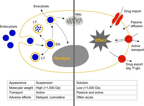
Figure 2 Known pathways of cellular uptake of nanoparticles.
Notes: Uptake of nanoparticles by eukaryotic cells is an active process. Endocytotic pathways include phagocytosis, and pinocytosis.
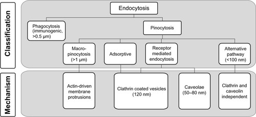
Figure 3 Experimental challenges and hurdles.
Notes: Specific physicochemical properties of ENMs may lead to technical challenges and artifacts in experimental systems. Particle agglomeration (A) reduces dosing accuracy or may lead to embolism after IV injection. Plasma-protein binding and opsonization of nanoparticles (B) may trigger a humoral immune response. Interaction of nanoparticles with cells of the MPS leads to accelerated plasma clearance (C). Accumulation of particles at a defined target site (D) might be impeded by their premature degradation and elimination (E).
Abbreviations: ENMs, engineered nanomaterials; IV, intravenous; MPS, mononuclear phagocytic system.
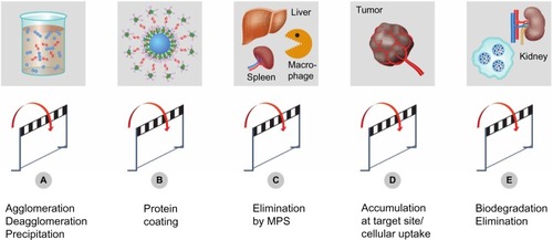
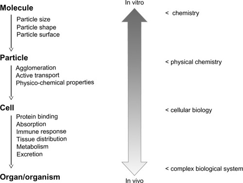
Figure 4 Extrapolation from in vitro data to the in vivo situation.
Notes: In vitro experimental systems can be used to characterize nanoparticles with respect to their chemical composition and physicochemical properties. Cell-culture-based experimental systems can be used to study molecular mechanisms of cellular uptake and intracellular processing of particles. However, additional information is needed to address questions related to the in vivo behavior of nanomaterials and their interaction with complex biological systems. In particular, the prediction of pharmacokinetic parameters remains a challenge.