Figures & data
Figure 1 Electron micrograph of nanoferromagnetic particles.
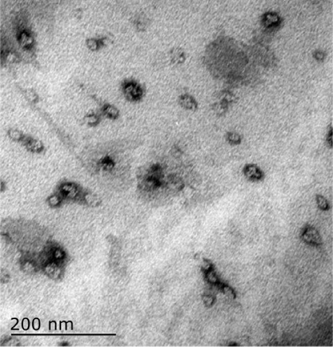
Figure 2 Time-temperature curve under an alternating magnetic field in vitro (Fe3O4, 20%, 2 mL).
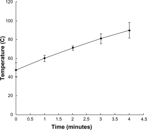
Figure 3 Angiography of the hepatic artery proper shows a clear rich blood supply to the tumor, especially at the rim.
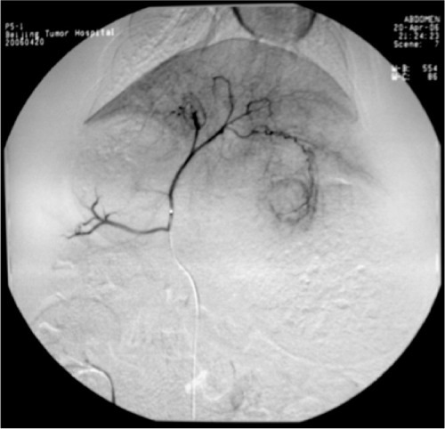
Figure 4 Embolization with magnetic particles and Lipiodol®, with the agent deposited well in the lesion, and quite a few particles seen in the normal hepatic parenchyma.
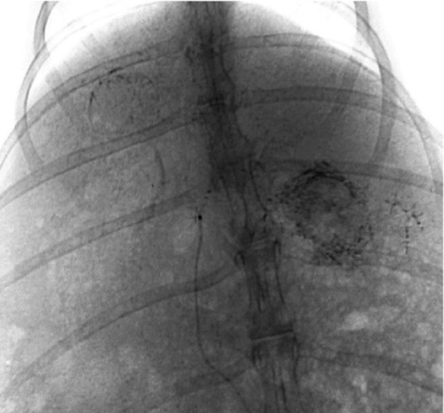
Figure 5 Perforations on stomach because of reflux of the mixed embolization agent.
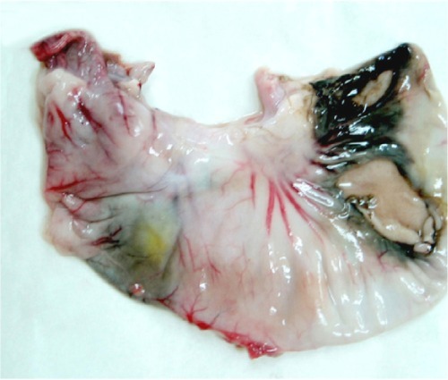
Table 1 Temperature at different tissue sites and times in groups B and D under an alternating magnetic field (°C)
Figure 6 Time-temperature curve for the three liver sites in group D in an alternating magnetic field.
Abbreviation: NHP, normal hepatic parenchyma.
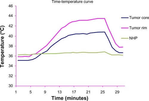
Figure 7 Adjusted by basal temperature, time-temperature curve at the three liver sites in group D, in an alternating magnetic field.
Abbreviation: NHP, normal hepatic parenchyma.
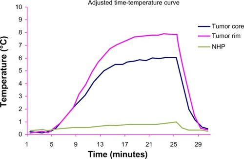
Table 2 Liver and kidney function in the four groups before and after treatment
Figure 8 Tumor section: inside and surrounding the tumor deposited a lot of black particles after magnetic embolization.
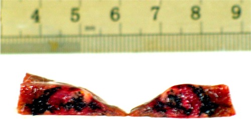
Table 3 Longest tumor diameter and volume of tumor before and after treatment
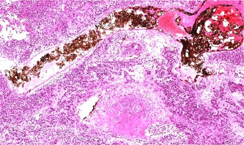
Figure 9 Light microscope: small artery surrounding tumor filled with black particles, tumor cell around necrosis (he, ×200).