Figures & data
Figure 1 (A) The synthesis of poly(ethylene glycol)-block-poly(D,L-lactic acid) (PEG1k-PDLLA10k) and (B) proton nuclear magnetic resonance spectra of mPEG1k-PDLLA in CDCl3 at 25°C. Characteristic peaks of PEG and PDLLA were located at about 3.6 ppm and 5.1–5.3 ppm (CH) and 1.5–1.6 ppm (CH3), respectively.
Abbreviation: Peg1k-PDLLA10k, polyethylene glycol-polylactide.
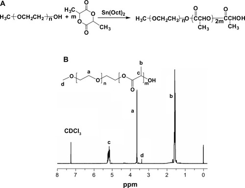
Figure 2 Synthesis of polymeric nanovesicles.
Abbreviations: DOX, doxorubicin; PDLLA, poly(D,L-lactic acid); PEG, poly(ethylene glycol); THF, tetrahydrofuran.
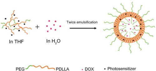
Figure 3 Transmission electron microscope photos of poly(ethylene glycol)-block-poly(D,L-lactic acid) vesicles. The structure of synthesized nanovesicles was clear, which was hollow and spherical. Particle size distribution was relatively homogeneous.
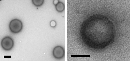
Figure 4 Particle sizing distribution of (A) poly(ethylene glycol)-block-poly(D,L-lactic acid) (PEG-PDLLA) blank vesicle, (B) doxorubicin (DOX)-loaded PEG-PDLLA vesicle, (C) photosensitizer-loaded PEG-PDLLA vesicle, and (D) DOX- and photosensitizer-loaded PEG-PDLLA vesicle.
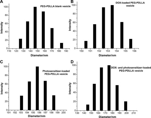
Figure 5 The toxicity of empty nanovesicles poly(ethylene glycol)-block-poly(D,L-lactic acid) (PEG-PDLLA) on HepG2.
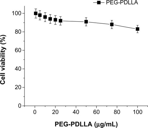
Figure 6 The toxicity of three groups of drug-loading nanoparticle vesicles.
Abbreviations: DOX, doxorubicin; HMME, hematoporphyrin monomethyl ether; PDLLA, poly(D,L-lactic acid); PEG, poly(ethylene glycol).
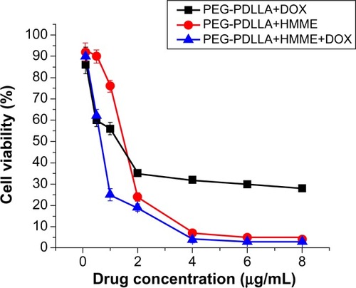
Figure 7 Reactive oxygen species levels in HepG2 cells following treatment with drug-loaded nanovesicles.
Abbreviations: DOX, doxorubicin; HMME, hematoporphyrin monomethyl ether; PDLLA, poly(D,L-lactic acid); PEG, poly(ethylene glycol).
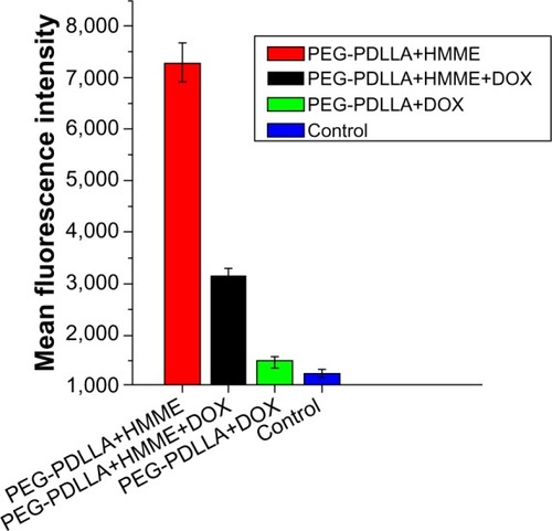
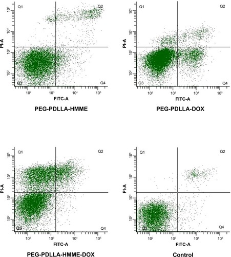
Figure 8 Mitochondrial membrane potential depolarization in HepG2 cells following treatment with drug-loaded nanovesicles.
Abbreviations: DOX, doxorubicin; HMME, hematoporphyrin monomethyl ether; PDLLA, poly(D,L-lactic acid); PEG, poly(ethylene glycol); FITC-A, fluorescein isothiocyanate apoptosis; PI-A, propidine iodide apoptosis; Q, quadrant.
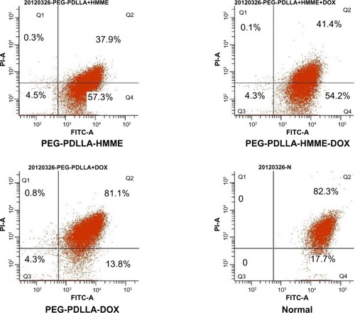
Figure 9 The apoptosis-inducing rate test result of drug-loaded nanovesicles.
Abbreviations: DOX, doxorubicin; HMME, hematoporphyrin monomethyl ether; PDLLA, poly(D,L-lactic acid); PEG, poly(ethylene glycol); FITC-A, fluorescein isothiocyanate apoptosis; PI-A, propidine iodide apoptosis; Q, quadrant.