Figures & data
Figure 1 (A) Endobronchial ultrasound tip with the ultrasound balloon dilated with water for injection. The biopsy tip is out of the plastic protection probe. (B) Lymph node seen on endobronchial ultrasound, showing the biopsy tip inside the lymph node. (C) Biopsy forceps and (D) biopsy needle.
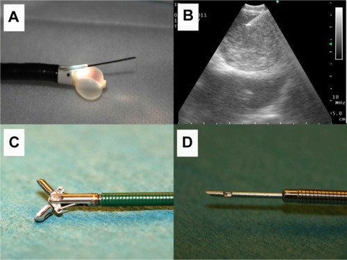
Figure 2 (A) Olympus tower, ready to use for bronchoscopy, mini-probe ultrasound, or endobronchial ultrasound. (B) Bronchoscope and (C) the “Stop effect” showing a tumor by mini-probe ultrasound. The Olympus ultrasound tip stops as it meets tumor tissue inside subsegmental alveolar tissue. (D) Biopsy forceps with tissue material and (E) Aspiration probe.
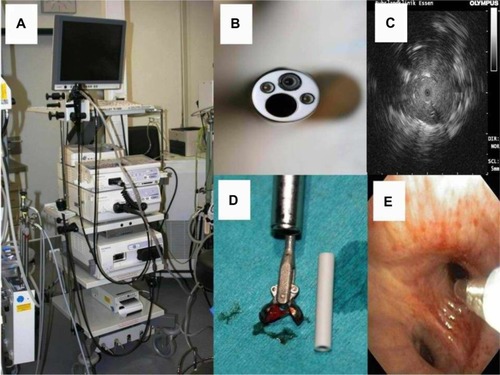
Figure 3 (A) Adenocarcinoma (microscopic observation 100×). (B) Adenocarcinoma with epidermal growth factor receptor mutation (microscopic observation 100×). (C) Squamous cell carcinoma (microscopic observation 100×). CytoViva® spectral imaging of (D) an adenocarcinoma, (E) an adenocarcinoma with epidermal growth factor receptor mutation, and (F) a squamous cell carcinoma.
Figure 7 (A) Cryptogenic organizing pneumonia (microscopic observation 100×). (B) Sarcoidosis (microscopic observation 100×). (C) Interstitial pulmonary fibrosis (microscopic observation 100×). CytoViva® spectral imaging of (D) cryptogenic organizing pneumonia, (E) sarcoidosis, and (F) interstitial pulmonary fibrosis.
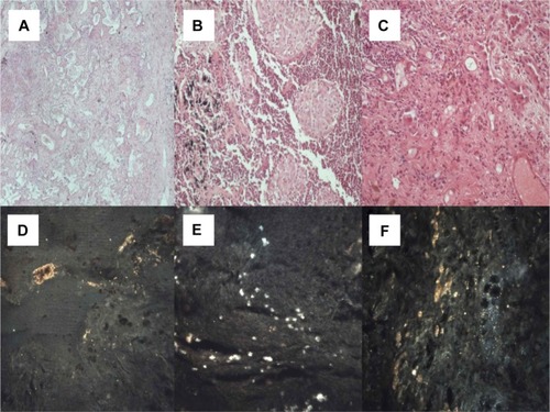
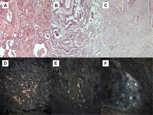
Figure 7 (A) Cryptogenic organizing pneumonia (microscopic observation 100×). (B) Sarcoidosis (microscopic observation 100×). (C) Interstitial pulmonary fibrosis (microscopic observation 100×). CytoViva® spectral imaging of (D) cryptogenic organizing pneumonia, (E) sarcoidosis, and (F) interstitial pulmonary fibrosis.
Figure 4 (A) Small cell neuroendocrine carcinoma (microscopic observation 100×). (B) Typical carcinoid tumor (microscopic observation 100×). (C) Thymoma (microscopic observation 100×). CytoViva® spectral imaging of (D) a small cell neuroendocrine carcinoma, (E) a typical carcinoid tumor, and (F) a thymoma.
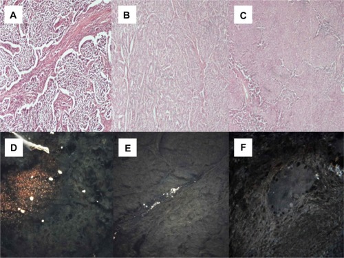
Figure 5 (A) Malt cell lymphoma (microscopic observation 100×). (B) Sarcomatoid mesothelioma (microscopic observation 100×). (C) Epithelioid mesothelioma (microscopic observation 100×). CytoViva® spectral imaging of (D) a malt cell lymphoma, (E) a sarcomatoid mesothelioma, and (F) an epithelioid mesothelioma.
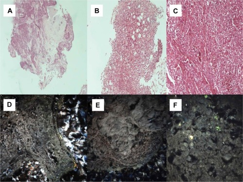
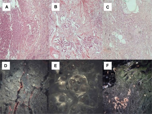
Figure 6 (A) Wegener’s granulomatosis (microscopic observation 100×). (B) Usual interstitial pneumonia (microscopic observation 100×). (C) Nonspecific interstitial pneumonia (microscopic observation 100 μm). (D) CytoViva® spectral imaging of Wegener’s granulomatosis, (E) usual interstitial pneumonia, and (F) nonspecific interstitial pneumonia.