Figures & data
Table 1 Phytochemical screening of leaf extract of Gymnema sylvestre
Table 2 Estimation of phytochemical compounds of leaf extract of Gymnema sylvestre
Figure 1 Surface plasmon resonance of silver nanoparticles.
Notes: (A) silver nitrate solution; (B) green-synthesized silver nanoparticles in ruby red color after 30 minutes.
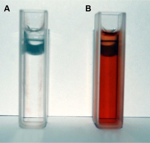
Figure 2 Time-dependent absorption spectra of silver nanoparticles after the bioreduction of silver in the aqueous extract of Gymnema sylvestre.
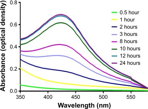
Figure 3 Scanning electron microscopic image of green silver nanoparticles synthesized by reduction of aqueous AgNO3 ions using aqueous extract of Gymnema sylvestre.
Abbreviations: HV, high voltage; WD, working distance; mag, magnification; ETD, Everhart–Thornley detector.
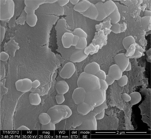
Figure 4 Energy-dispersive X-ray spectrum of green silver nanoparticles.
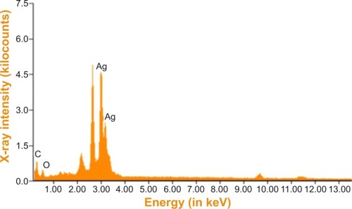
Figure 5 Fourier transform infrared spectroscopy spectrum of green-synthesized silver nanoparticles along with the plant extract of Gymnema sylvestre (GS).
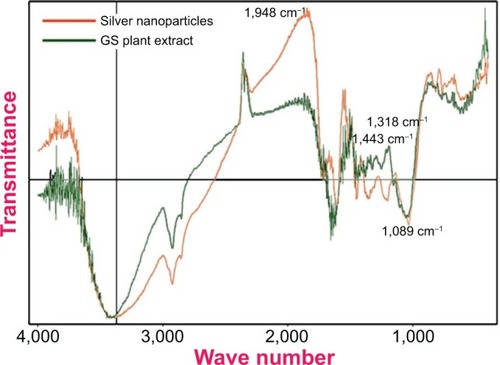
Figure 6 X-ray diffraction spectrum of green-synthesized silver nanoparticles.
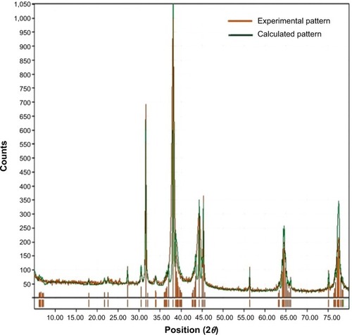
Table 3 Comparative chart of the characterization of green-synthesized silver nanoparticles using various plant extracts
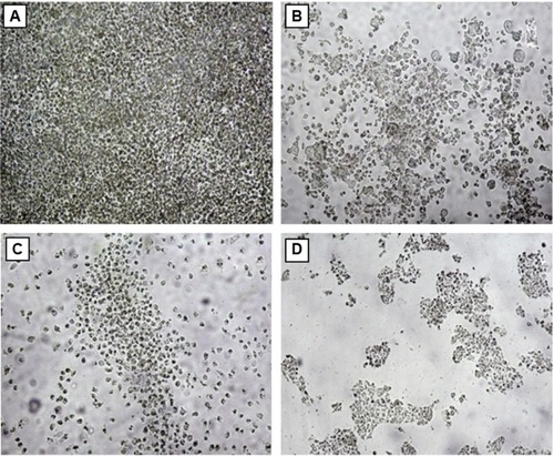
Figure 7 Anticancer activity of the green-synthesized silver nanoparticles.
Abbreviation: GS, Gymnema sylvestre.
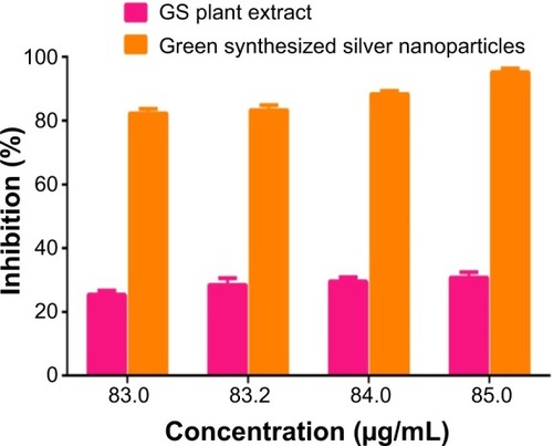
Figure 8 Inverted microscopic image of HT29 (A) control cells, (B) Gymnema sylvestre-treated (85 μg/mL), (C) silver nanoparticle-treated (83 μg/mL), and (D) silver nanoparticle-treated (85 μg/mL) cells.