Figures & data
Figure 1 Steps for preparation of the Cy5.5-Lf-SPIO micelles.
Abbreviations: Lf, lactoferrin; SPIO, superparamagnetic iron oxide; SIA, succinimidyl iodoacetate.
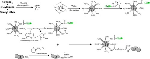
Figure 2 Morphology and size distribution of the materials.
Notes: (A) TEM image of SPIO. (B) TEM image of Cy5.5-Lf-SPIO micelles. (C) Hydrodynamic size distribution of Cy5.5-Lf-SPIO micelles at 25°C. (D) Long-term stability based on hydrodynamic size change in phosphate-buffered saline over time at room temperature.
Abbreviations: TEM, transmission electron microscopy; Lf, lactoferrin; SPIO, superparamagnetic iron oxide.
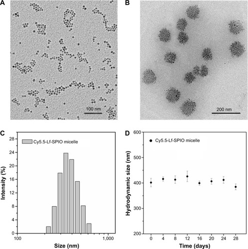
Figure 3 Magnetic properties and MR imaging abilities of the materials.
Notes: (A) Magnetization curves at 300 K. (B) T2-weighted MR images of the Cy5.5-Lf-SPIO micelles. (C) Relaxivities (r2) of the Cy5.5-Lf-SPIO micelles measured at 300 K.
Abbreviations: MR, magnetic resonance; Lf, lactoferrin; SPIO, superparamagnetic iron oxide.
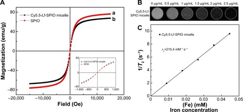
Figure 4 Fluorescence imaging abilities of the materials.
Notes: (A) Fluorescence spectra at the excitation wavelength of 640 nm at room temperature. (B) Fluorescent image of the Cy5.5-Lf-SPIO micelles. (C) The correlation between fluorescence intensity and the mass of the iron.
Abbreviations: Lf, lactoferrin; SPIO, superparamagnetic iron oxide.
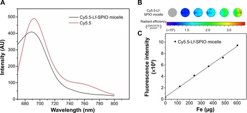
Figure 5 In vitro study of the materials.
Notes: (A) In vitro T2-weighted magnetic resonance images of C6 cells with different treatments. (B) Fluorescence images of C6 cells with different treatments. (C) Confocal fluorescence images of C6 cells with different treatments (400×).
Abbreviations: Lf, lactoferrin; SPIO, superparamagnetic iron oxide; DAPI, 4′,6-diamidino-2-phenylindole.
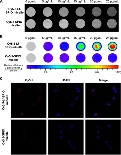
Figure 6 In vivo study on magnetic resonance imaging.
Notes: (A) In vivo T2-weighted magnetic resonance images of rat brain bearing C6 glioma (n=6). Upper row, acquired after administration of Cy5.5-Lf-SPIO micelles (12 mg Fe/kg); lower row, acquired after administration of Cy5.5-SPIO micelles (12 mg Fe/kg). (B) The relative signal enhancement of the brain tumor in the T2-weighted image at 24 and 48 hours (expressed as means ± standard error of mean [n=6]). (C) Histological sections of glioma with Prussian blue staining (400×).
Abbreviations: Lf, lactoferrin; SPIO, superparamagnetic iron oxide.
![Figure 6 In vivo study on magnetic resonance imaging.Notes: (A) In vivo T2-weighted magnetic resonance images of rat brain bearing C6 glioma (n=6). Upper row, acquired after administration of Cy5.5-Lf-SPIO micelles (12 mg Fe/kg); lower row, acquired after administration of Cy5.5-SPIO micelles (12 mg Fe/kg). (B) The relative signal enhancement of the brain tumor in the T2-weighted image at 24 and 48 hours (expressed as means ± standard error of mean [n=6]). (C) Histological sections of glioma with Prussian blue staining (400×).Abbreviations: Lf, lactoferrin; SPIO, superparamagnetic iron oxide.](/cms/asset/7a3109c9-4caa-43fe-86ba-6cf1b4cb6be3/dijn_a_72910_f0006_c.jpg)
Figure 7 Ex vivo study of the brain tissue.
Notes: (A) Ex vivo fluorescence images and H&E-staining images of rat brain bearing C6 glioma at 48 hours postinjection. (B) The average fluorescence intensity of the brain tumor and the normal brain. Results expressed as means ± standard error of mean (n=6).
Abbreviations: Lf, lactoferrin; SPIO, superparamagnetic iron oxide; H&E, hematoxylin and eosin.
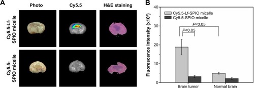
Figure 8 The confocal fluorescence images of the brain slices.
Notes: Upper row, sections of the group with treatment of Cy5.5-Lf-SPIO micelles (100×); lower row, sections of the group with treatment of Cy5.5-SPIO micelles (100×).
Abbreviations: Lf, lactoferrin; SPIO, superparamagnetic iron oxide; DAPI, 4′,6-diamidino-2-phenylindole.
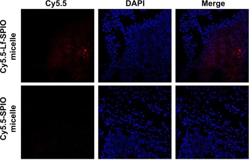
Figure 9 Viabilities of NIH-3T3 cells treated with Cy5.5-Lf-SPIO micelles in cytotoxicity studies. Results expressed as means ± standard error of mean.
Abbreviations: Lf, lactoferrin; SPIO, superparamagnetic iron oxide.
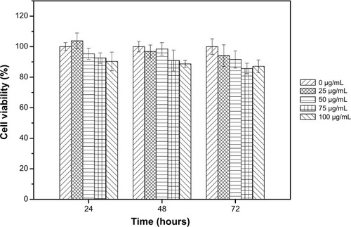
Figure 10 Illustration of the multiple functions of Cy5.5-Lf-SPIO micelles.
Abbreviations: Lf, lactoferrin; SPIO, superparamagnetic iron oxide; PEG-b-PCL, polyethylene glycol-block-polycaprolactone; MR, magnetic resonance.
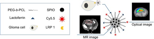
Figure S1 Fourier-transform infrared spectra of SPIO micelles, lactoferrin, and Cy5.5-Lf-SPIO micelle.
Abbreviation: SPIO, superparamagnetic iron oxide.
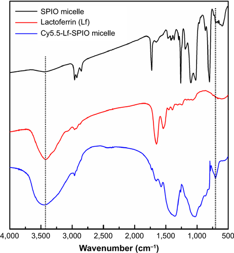
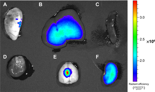
Figure S2 Optical images of rat organs.
Notes: (A) Lung; (B) liver; (C) spleen; (D) heart; (E) brain; (F) kidney.