
Figures & data
Figure 1 Effect of glycosaminoglycan (GAG) concentration on poly-lactide-co-glycolide (PLGA) nanoparticle (NP) size (n=3): (A) “0.00 mg/mL” corresponds to plain PLGA NPs; (B) transmission electron micrographs of GAG-functionalized PLGA NPs.
Abbreviations: PLGA/CS, poly-lactide-co-glycolide functionalized with chondroitin sulfate; PLGA/DS, poly-lactide-co-glycolide functionalized with dermatan sulfate; PLGA/H, poly-lactide-co-glycolide functionalized with heparin; PLGA/HA, poly-lactide-co-glycolide functionalized with hyaluronic acid.
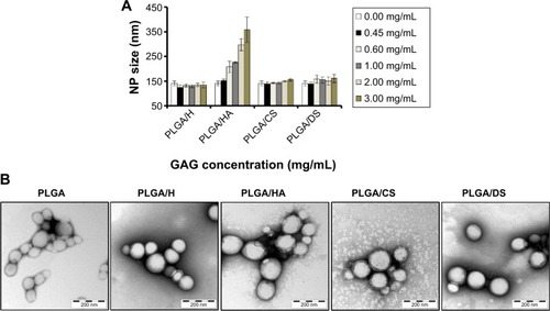
Table 1 Surface characteristics of glycosaminoglycan-functionalized poly-lactide-co-glycolide (PLGA) nanoparticles (NPs)
Figure 2 (A) Zeta potential measurement of glycosaminoglycan (GAG)-functionalized poly-lactide-co-glycolide (PLGA) nanoparticles (NPs) in deionized water, cell-culture medium, and phosphate-buffered saline (pH 7.4) (n=3). (B) Isoelectric point measurement of the GAG-functionalized PLGA NPs as a function of pH.
Abbreviations: PLGA/CS, poly-lactide-co-glycolide functionalized with chondroitin sulfate; PLGA/DS, poly-lactide-co-glycolide functionalized with dermatan sulfate; PLGA/H, poly-lactide-co-glycolide functionalized with heparin; PLGA/HA, poly-lactide-co-glycolide functionalized with hyaluronic acid.
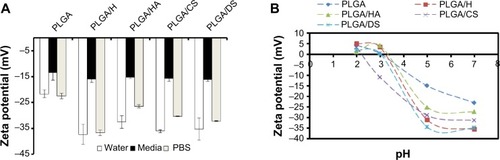
Table 2 Elemental composition of glycosaminoglycan-functionalized poly-lactide-co-glycolide (PLGA) nanoparticles (NPs) determined using X-ray photon electron spectroscopy
Figure 3 Cumulative carbon 1s (C1s) X-ray photoelectron spectra of poly-lactide-co-glycolide (PLGA) nanoparticles bearing different glycosaminoglycans demonstrating binding energies of different glycosaminoglycans. C1s peaks at 289.1 eV, 287 eV and 285.1 eV correspond to O-C=O, C-O-C and C-H and C-C bonds respectively in all samples. C1s peaks at 285 eV and a shoulder at 285.5 eV correspond to C-S and C-N in PLGA/H, PLGA/CS and PLGA/DS and correspond to C-N in PLGA/HA.
Notes: X-ray photoelectron spectra of PLGA NPs: (A), PLGA; (B), PLGA/H; (C), PLGA/HA; (D), PLGA/CS; (E), PLGA/DS.
Abbreviations: PLGA/CS, poly-lactide-co-glycolide functionalized with chondroitin sulfate; PLGA/DS, poly-lactide-co-glycolide functionalized with dermatan sulfate; PLGA/H, poly-lactide-co-glycolide functionalized with heparin; PLGA/HA, poly-lactide-co-glycolide functionalized with hyaluronic acid.
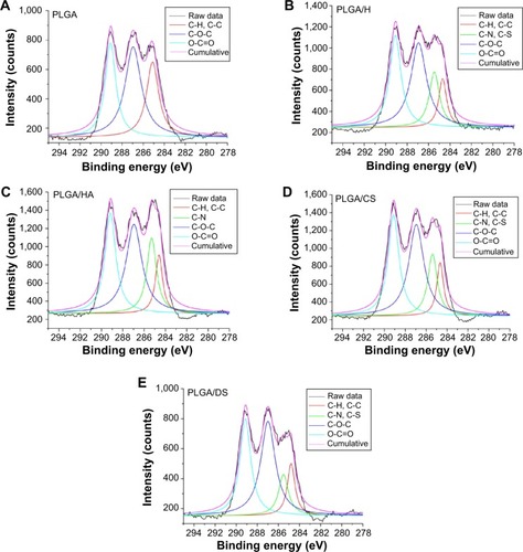
Figure 4 Viability (as a percentage of tissue-culture plastic control) of (A) lung epithelial adenocarcinoma (A549) cells, (B) human pulmonary microvascular endothelial cells (HPMEC), and (C) renal proximal tubular epithelial cells (RPTEC) following exposure to increasing concentrations of glycosaminoglycan-functionalized poly-lactide-co-glycolide (PLGA) nanoparticles (NPs) for 24 hours (n=3).
Abbreviations: PLGA/CS, poly-lactide-co-glycolide functionalized with chondroitin sulfate; PLGA/DS, poly-lactide-co-glycolide functionalized with dermatan sulfate; PLGA/H, poly-lactide-co-glycolide functionalized with heparin; PLGA/HA, poly-lactide-co-glycolide functionalized with hyaluronic acid.
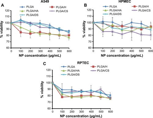
Figure 5 Effect of glycosaminoglycan-functionalized poly-lactide-co-glycolide (PLGA) nanoparticles (NPs) on cell-membrane integrity following exposure to increasing concentrations of GAG-functionalized PLGA NPs for 24 hours. The relative lactate dehydrogenase (LDH) release was co-related to percentage cytotoxicity. Cytotoxicity due to loss in membrane integrity in (A) lung epithelial adenocarcinoma (A549) cells, (B) human pulmonary microvascular endothelial cells (HPMEC), and (C) renal proximal tubular epithelial cells (RPTEC) (n=3).
Abbreviations: PLGA/CS, poly-lactide-co-glycolide functionalized with chondroitin sulfate; PLGA/DS, poly-lactide-co-glycolide functionalized with dermatan sulfate; PLGA/H, poly-lactide-co-glycolide functionalized with heparin; PLGA/HA, poly-lactide-co-glycolide functionalized with hyaluronic acid.
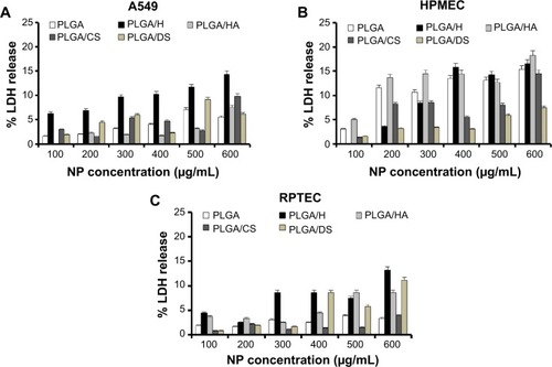
Figure 6 Reactive oxygen species induction in (A) lung epithelial adenocarcinoma (A549) cells, (B) human pulmonary microvascular endothelial cells (HPMEC), and (C) renal proximal tubular epithelial cells (RPTEC), following incubation with glycosaminoglycan-functionalized poly-lactide-co-glycolide (PLGA) nanoparticles (NPs) for 24 hours (n=3).
Abbreviations: DCF, dichlorofluorescein; PLGA/CS, poly-lactide-co-glycolide functionalized with chondroitin sulfate; PLGA/DS, poly-lactide-co-glycolide functionalized with dermatan sulfate; PLGA/H, poly-lactide-co-glycolide functionalized with heparin; PLGA/HA, poly-lactide-co-glycolide functionalized with hyaluronic acid.
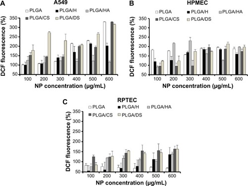
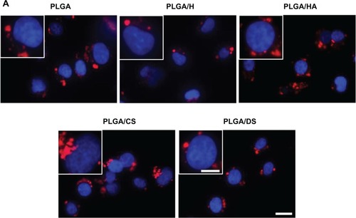
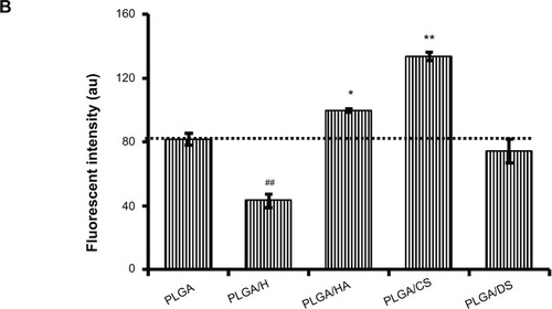
Figure 7 Fluorescent micrographs (A) and fluorescence-activated cell-sorting histogram (B) demonstrating uptake of glycosaminoglycan-functionalized poly-lactide-co-glycolide (PLGA) nanoparticles by lung epithelial adenocarcinoma (A549) cells after 4 hours of incubation (n=3).
Figure 8 Fluorescent micrographs (A) and fluorescence-activated cell-sorting histogram (B) demonstrating uptake of glycosaminoglycan-functionalized poly-lactide-co-glycolide (PLGA) nanoparticles (NPs) by human pulmonary microvascular endothelial cells (HPMEC) after 4 hours of incubation (n=3).
Notes: *P<0.05 between PLGA and PLGA/HA. **P<0.005 between PLGA and PLGA/CS. ##P<0.005 between PLGA and PLGA/H. Scale bar: 20 μm for low magnification and 10 μm for high magnification (inset).
Abbreviations: PLGA/CS, poly-lactide-co-glycolide functionalized with chondroitin sulfate; PLGA/DS, poly-lactide-co-glycolide functionalized with dermatan sulfate; PLGA/H, poly-lactide-co-glycolide functionalized with heparin; PLGA/HA, poly-lactide-co-glycolide functionalized with hyaluronic acid.
Notes: *P<0.05 between PLGA and PLGA/HA. **P<0.005 between PLGA and PLGA/CS. ##P<0.005 between PLGA and PLGA/DS. ###P<0.0005 between PLGA and PLGA/H. Scale bar: 20 μm for low magnification and 10 μm for high magnification (inset).
Abbreviations: DCF, dichlorofluorescein; PLGA/CS, poly-lactide-co-glycolide functionalized with chondroitin sulfate; PLGA/DS, poly-lactide-co-glycolide functionalized with dermatan sulfate; PLGA/H, poly-lactide-co-glycolide functionalized with heparin; PLGA/HA, poly-lactide-co-glycolide functionalized with hyaluronic acid.
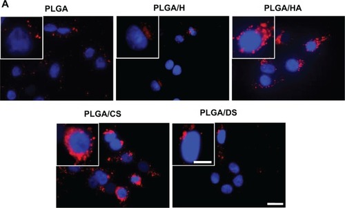
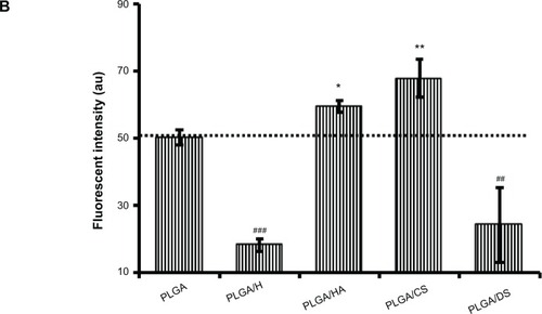
Figure 8 Fluorescent micrographs (A) and fluorescence-activated cell-sorting histogram (B) demonstrating uptake of glycosaminoglycan-functionalized poly-lactide-co-glycolide (PLGA) nanoparticles (NPs) by human pulmonary microvascular endothelial cells (HPMEC) after 4 hours of incubation (n=3).
Notes: *P<0.05 between PLGA and PLGA/HA. **P<0.005 between PLGA and PLGA/CS. ##P<0.005 between PLGA and PLGA/H. Scale bar: 20 μm for low magnification and 10 μm for high magnification (inset).
Abbreviations: PLGA/CS, poly-lactide-co-glycolide functionalized with chondroitin sulfate; PLGA/DS, poly-lactide-co-glycolide functionalized with dermatan sulfate; PLGA/H, poly-lactide-co-glycolide functionalized with heparin; PLGA/HA, poly-lactide-co-glycolide functionalized with hyaluronic acid.
Figure S1 Uptake of glycosaminoglycan-functionalized poly-lactide-co-glycolide (PLGA) nanoparticles by lung epithelial adenocarcinoma (A549) cells after 4 hours of incubation as depicted by overlay of differential interference contrast and fluorescence images.
Note: Scale bar: 20 μm.
Abbreviations: PLGA/CS, poly-lactide-co-glycolide functionalized with chondroitin sulfate; PLGA/DS, poly-lactide-co-glycolide functionalized with dermatan sulfate; PLGA/H, poly-lactide-co-glycolide functionalized with heparin; PLGA/HA, poly-lactide-co-glycolide functionalized with hyaluronic acid.
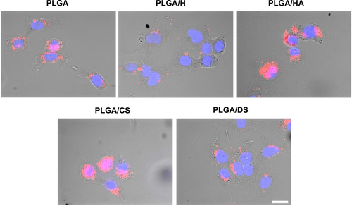
Figure S2 Uptake of glycosaminoglycan-functionalized poly-lactide-co-glycolide (PLGA) nanoparticles by human pulmonary microvascular endothelial cells after 4 hours of incubation as depicted by overlay of differential interference contrast and fluorescence images.
Note: Scale bar: 20 μm.
Abbreviations: PLGA/CS, poly-lactide-co-glycolide functionalized with chondroitin sulfate; PLGA/DS, poly-lactide-co-glycolide functionalized with dermatan sulfate; PLGA/H, poly-lactide-co-glycolide functionalized with heparin; PLGA/HA, poly-lactide-co-glycolide functionalized with hyaluronic acid.
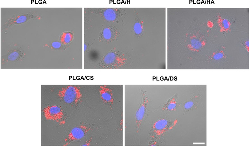
Table S1 Size distribution of glycosaminoglycan (GAG)-functionalized poly-lactide-co-glycolide (PLGA) nanoparticles (NPs) (0.45 mg/mL of GAG) as determined by dynamic light scattering
Table S2 Size distribution of glycosaminoglycan (GAG)-functionalized poly-lactide-co-glycolide (PLGA) nanoparticles (NPs) (0.45 mg/mL of GAG) as determined by transmission electron microscopy
Table S3 Binding energy of functional groups of glycosaminoglycan-functionalized poly-lactide-co-glycolide (PLGA) nanoparticles as determined by X-ray photoelectron spectroscopy