Figures & data
Figure 1 Characterization of SCs.
Notes: Double-immunofluorescent staining showed the expression of S-100 (A and E) and Sox-10 (B and F) with DAPI nuclear counterstaining (C and G). Merge image showed a purity of more than 96% SCs (D and H). Scale bars: (A–D) 100 µm (magnification 20×), (E–H) 50 µm (magnification 40×).
Abbreviations: SCs, Schwann cells; DAPI, 4′,6-diamidino-2-phenylindole.
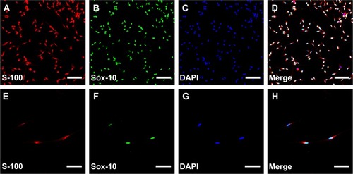
Figure 2 Schematic view of this experiment.
Notes: (A) Experimental time scale. (B) The device used to expose SCs to the MF and the no-MF stimulations. The solenoid was wired to a pulse generator. (C) The membrane and scaffold seeded with SC model. (D) The distribution of the MF in the solenoid. Models were placed in the axial plane of the solenoid.
Abbreviations: SCs, Schwann cells; MF, magnetic field.
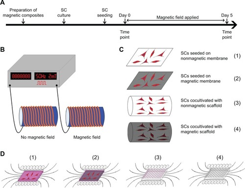
Table 1 Primer sequences used for real-time polymerase chain reaction
Figure 3 Characterization of MNPs.
Notes: (A) TEM. (B) Size distribution of MNPs. (C) XRD. (D) FTIR spectrum. (E) Magnetization of MNPs.
Abbreviations: MNPs, magnetic nanoparticles; TEM, transmission electron microscopy; XRD, X-ray diffraction; FTIR, Fourier-transform infrared.
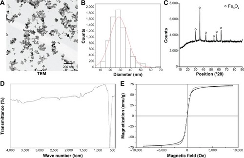
Figure 4 FTIR spectrum and XRD images of the magnetic nanocomposites.
Notes: The FTIR (A) and XRD (B) spectra of all the magnetic nanocomposites were similar, although they differed slightly from each other due to different weight ratios of MNPs.
Abbreviations: FTIR, Fourier-transform infrared; XRD, X-ray diffraction; MNPs, magnetic nanoparticles.
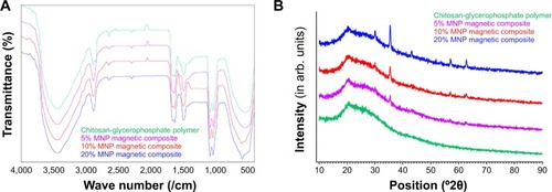
Figure 5 The appearance and magnetization of magnetic scaffolds.
Notes: (A) The appearance of magnetic scaffolds. From left to right: nonmagnetic scaffold, 5% MNP magnetic scaffold, 10% MNP magnetic scaffold, and 20% MNP magnetic scaffold. (B) The magnetization of magnetic nanocomposites was tested with a VSM. Photographs showing the attraction of the 10% MNP magnetic scaffold to a standard magnet after magnetization: up–down (C), and in–out (D).
Abbreviations: MNP, magnetic nanoparticle; VSM, vibrating sample magnetometer.
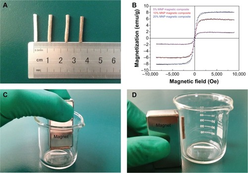
Figure 6 SEM images of the magnetic membranes and scaffolds.
Notes: (A–D) Representative images of nonmagnetic membrane, 5% MNP magnetic membrane, 10% MNP magnetic membrane, and 20% MNP magnetic membrane are shown in the first row. (E–L) Representative images of nonmagnetic scaffold, 5% MNP magnetic scaffold, 10% MNP magnetic scaffold, and 20% MNP magnetic scaffold in a longitudinal section, showing the longitudinally oriented microchannels and interconnected porous structure. (M–P) Representative images of nonmagnetic scaffold, 5% MNP magnetic scaffold, 10% MNP magnetic scaffold, and 20% MNP magnetic scaffold in a transverse section, showing that the microchannels were arranged in a honeycomb-like pattern. Scale bars: (A–D) 1.5 µm, (E–H) 60 µm, (I–P) 30 µm.
Abbreviations: SEM, scanning electron microscopy; MNPs, magnetic nanoparticles.
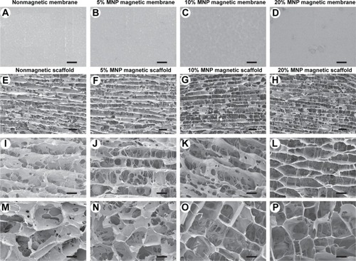
Figure 7 Apoptosis assay of SCs in each group by flow cytometry and CCK-8 assay.
Notes: (A) Culture-plate group; (B) nonmagnetic membrane group; (C) magnetic membrane group; (D) magnetic membrane + MF group (0.5 mT); (E) magnetic membrane + MF group (1.0 mT); (F) nonmagnetic membrane + MF group (2.0 mT); (G) magnetic membrane + MF group (2.0 mT); (H) nonmagnetic membrane + MF group (5.0 mT); (I) magnetic membrane + MF group (5.0 mT); (J) nonmagnetic membrane + MF group (10.0 mT); (K) magnetic membrane + MF group (10.0 mT). (L) The percentage of apoptotic cells in each group was obtained by averaging the results of five flow-cytometry assays for each group. (M) The CCK-8 values of viable cells in each group were obtained. All data are expressed as means ± standard error of mean. *P<0.05; **P<0.01, one-way ANOVA when compared with culture-plate group.
Abbreviations: SCs, Schwann cells; MF, magnetic field; CCK, Cell Counting Kit; ANOVA, analysis of variance; PI, propidium iodide; h, hours; NS, not significant.
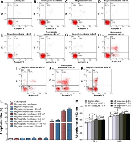
Figure 8 Representative SEM photomicrographs of SCs in each group at 24 hours after seeding.
Notes: (A) Culture-plate group; (B) nonmagnetic membrane group; (C) magnetic membrane group; (D) culture plate + MF group (2.0 mT); (E) nonmagnetic membrane + MF group (2.0 mT); (F) magnetic membrane + MF group (2.0 mT); (G) dividing phase in magnetic membrane + MF group (2.0 mT); (H) magnetic membrane + MF group (5.0 mT); (I) magnetic membrane + MF group (10.0 mT). (J) The dendritic length of SCs in each group was measured (n=20). Scale bar: (A, D, E, G) 20 µm, (B, F, I) 15 µm, (C) 10 µm, (H) 5 µm.
Abbreviations: SCs, Schwann cells; MF, magnetic field.
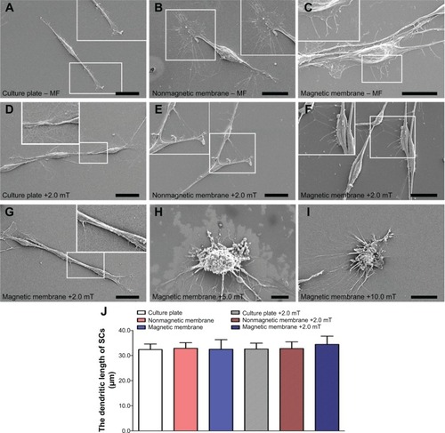
Figure 9 EdU staining and PrestoBlue assay in magnetic membranes at 24 hours after seeding.
Notes: (A) culture plate group; (B) nonmagnetic membrane group; (C) magnetic membrane group; (D) culture plate + MF group (2.0 mT); (E) nonmagnetic membrane + MF group (2.0 mT); (F) magnetic membrane + MF group (2.0 mT). The EdU-labeling index (G) and PrestoBlue assay values (H) in each group were obtained by averaging the results of five samples for each group. Scale bar: (A–F) 50 µm (magnification 20×). All data are expressed as means ± standard error of mean. **P<0.01.
Abbreviations: EdU, 5-ethynyl-2-deoxyuridine; MF, magnetic field; NS, not significant.
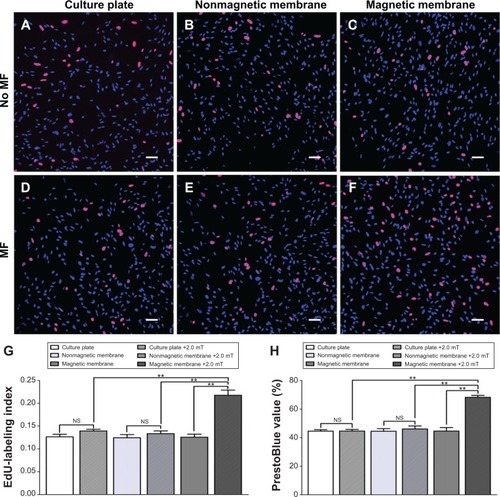
Figure 10 SEM and Live/Dead staining of the SCs cocultivated with scaffold in each group.
Notes: (A–D) Representative images of SEM at 24 hours after seeding, (E–H) Live/Dead staining at Day 1, (I–L) Day 3, and (M–P) Day 5 in the nonmagnetic scaffold group, nonmagnetic scaffold + MF group (2.0 mT), magnetic scaffold group, and magnetic scaffold + MF group (2.0 mT). The percentage of living SCs (Q) and PrestoBlue assay values (R) in each group were obtained by averaging the results of five samples for each group. Scale bars: (E–P) 50 µm (magnification 60×). All data are expressed as means ± standard error of mean. *P<0.05; **P<0.01.
Abbreviations: SEM, scanning electron microscopy; SCs, Schwann cells; MF, magnetic field; NS, not significant.
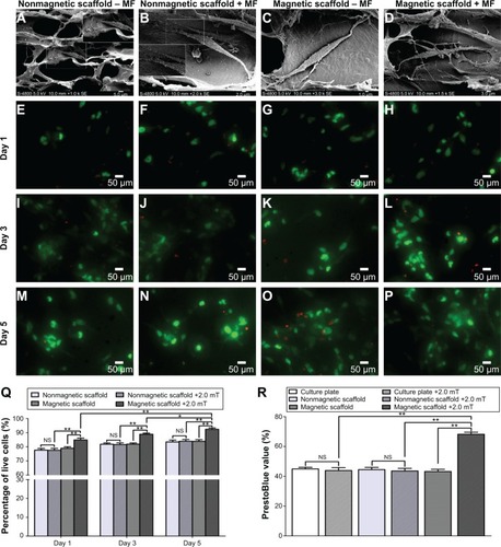
Figure 11 mRNA levels of BDNF, GDNF, NT-3, and VEGF in each group with or without MF at 12, 24, and 36 hours after MF.
Notes: mRNA levels of (A) BDNF, (B) GDNF, (C) NT-3, and (D) VEGF were determined for the culture-plate group, nonmagnetic membrane group, nonmagnetic membrane + MF group (2.0 mT), magnetic membrane group, magnetic membrane + MF group (2.0 mT), nonmagnetic scaffold group, nonmagnetic scaffold + MF group (2.0 mT), magnetic scaffold group, and magnetic scaffold + MF group (2.0 mT). Each test was repeated three times. The ratio of mRNA levels of the SC nonmagnetic membrane group, nonmagnetic membrane + MF group (2.0 mT), magnetic membrane group, magnetic membrane + MF group (2.0 mT), nonmagnetic scaffold group, nonmagnetic scaffold + MF group (2.0 mT), magnetic scaffold group, and magnetic scaffold + MF group (2.0 mT) to the culture plate group are shown. All data are expressed as means ± standard error of mean. **P<0.01.
Abbreviations: mRNA, messenger ribonucleic acid; BDNF, brain-derived neurotrophic factor; GDNF, glial cell-derived neurotrophic factor; NT-3, neurotrophin 3; VEGF, vascular endothelial growth factor; MF, magnetic field; SC, Schwann cell; NS, not significant; h, hours.
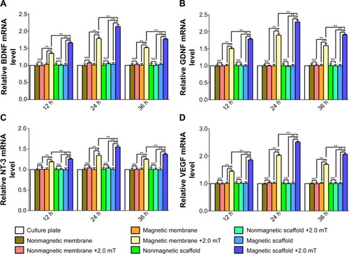
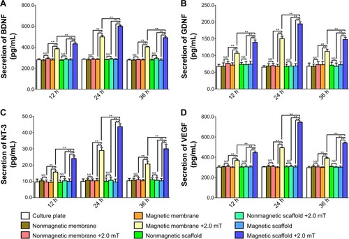
Figure 12 Secretion of BDNF, GDNF, NT-3, and VEGF in each group with or without MF at 12, 24, and 36 hours after MF.
Notes: Secretion levels of (A) BDNF, (B) GDNF, (C) NT-3, and (D) VEGF were determined for the culture-plate group, nonmagnetic membrane group, nonmagnetic membrane + MF group (2.0 mT), magnetic membrane group, magnetic membrane + MF group (2.0 mT), nonmagnetic scaffold group, nonmagnetic scaffold + MF group (2.0 mT), magnetic scaffold group, and magnetic scaffold + MF group (2.0 mT). Each test was repeated three times. Protein-level ratios of the SC nonmagnetic membrane group, nonmagnetic membrane + MF group (2.0 mT), magnetic membrane group, magnetic membrane + MF group (2.0 mT), nonmagnetic scaffold group, nonmagnetic scaffold + MF group (2.0 mT), magnetic scaffold group, and magnetic scaffold + MF group (2.0 mT) to the culture-plate group are shown. All data are expressed as means ± standard error of mean. **P<0.01.
Abbreviations: BDNF, brain-derived neurotrophic factor; GDNF, glial cell-derived neurotrophic factor; NT-3, neurotrophin 3; VEGF, vascular endothelial growth factor; MF, magnetic field; SC, Schwann cell; NS, not significant; h, hours.