Figures & data
Figure 1 Scanning electron microscopy images of hollow hydroxyapatite microspheres.
Notes: (A) External surface under low magnification. (B) Fractured section under high magnification.
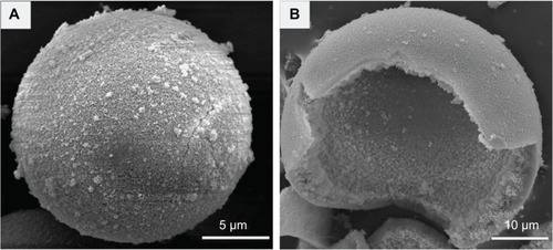
Figure 2 Cumulative release of recombinant human bone morphogenetic protein 2 (rhBMP2) from hollow hydroxyapatite microspheres into phosphate-buffered saline.
Note: The insert graph shows an initial burst release within 48 hours.
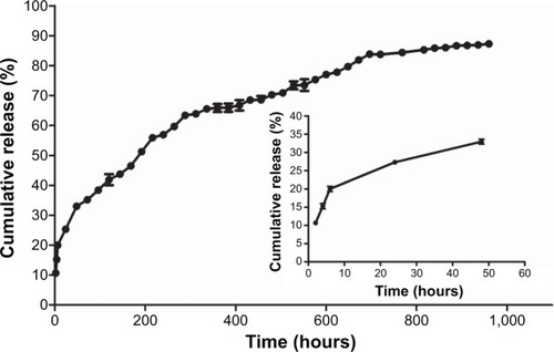
Figure 3 Serum alkaline phosphatase level.
Notes: Group A: BMP2-loaded hollow HA microspheres; Group B: hollow HA microspheres without BMP2; Group C: soluble BMP2 without a carrier. Different symbols (&, #, and *) represent different serum alkaline phosphatase activities at each time point (P<0.05). N=4/group/time point.
Abbreviations: HA, hydroxyapatite; BMP2, bone morphogenetic protein 2; S-ALP, serum alkaline phosphatase.
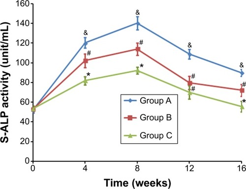
Figure 4 X-ray images of segmental radius at 4, 8, 12, and 16 weeks with different implants.
Notes: (A) BMP2-loaded hollow HA microspheres; (B) hollow HA microspheres without BMP2; (C) soluble BMP2 without a carrier. Arrows: defective and repairing sites.
Abbreviations: HA, hydroxyapatite; BMP2, bone morphogenetic protein 2.
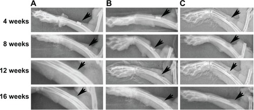
Table 1 Lane–Sandhu radiographical bone formation score (mean ± standard deviation)
Figure 5 Three-dimensional tomographic images of the segmental radius at 4 and 16 weeks.
Notes: (A) BMP2-loaded hollow HA microspheres; (B) hollow HA microspheres without BMP2; (C) soluble BMP2 without a carrier.
Abbreviations: HA, hydroxyapatite; BMP2, bone morphogenetic protein 2.
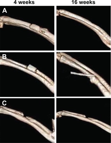
Table 2 Percentage of new bone formation (mean ± standard deviation)
Figure 6 Histological evaluation of three groups at 4, 8, 12, and 16 weeks.
Notes: (A) BMP2-loaded hollow HA microspheres; (B) hollow HA microspheres without BMP2; (C) soluble BMP2 without a carrier. Scale bars: 400 μm, 10× magnification; 100 μm, 40× magnification.
Abbreviations: BC, blood cells; BMP2, bone morphogenetic protein 2; CC, chondrocytes; FT, fibrous tissue; HA, hydroxyapatite; IC, inflammatory cells; Mat, material; NB, new bone; NLB, new lamellar bone; OB, osteoblasts; OC, osteocytes; OT, osteon; V, vessel.
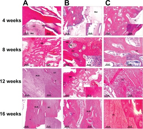
Figure 7 Counts from ROI at the fractured side.
Notes: (A) Blood pool phase; (B) static phase. Group A: BMP2-loaded hollow HA microspheres; Group B hollow HA microspheres without BMP2; Group C soluble BMP2 without a carrier. Different symbols (&, # and *) represent different ROI counts at each time point (P<0.05). N=4/group/time point.
Abbreviations: BMP2, bone morphogenetic protein 2; HA, hydroxyapatite; ROI, region of interest.
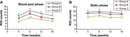
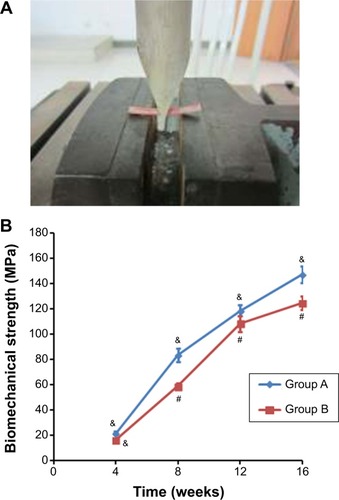
Figure 8 Biomechanical test of defective radius.
Notes: (A) mechanical testing facility; (B) the maximum flexural strength of defective radius. Group A: BMP2-loaded hollow HA microspheres; Group B, hollow HA microspheres without BMP2. Different symbols (& and #) represent different biomechanical strengths at each time point (P<0.05). N=4/group/time point.
Abbreviations: BMP2, bone morphogenetic protein 2; HA, hydroxyapatite.