Figures & data
Figure 1 Preparation scheme of the Qu-M via a self-assembly method.
Notes: First, quercetin (A) and MPEG–PCL (B) were codissolved in the acetone solution. In this process, quercetin and MPEG–PCL were assembled automatically into the Qu-M (C).
Abbreviations: PCL, poly(ε-caprolactone); PEG, poly(ethylene glycol); QU, quercetin; Qu-M, quercetin-loaded MPEG–PCL nanomicelles; MPEG–PCL, monomethoxy poly(ethylene glycol)–poly(ε-caprolactone).
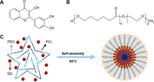
Figure 2 Characterizations of the Qu-M.
Notes: (A) Size distribution of Qu-M, (B) zeta potential of Qu-M, (C) TEM image of Qu-M, (D) PBS solution, Qu-M in PBS solution, and free quercetin in PBS solution (from left to right).
Abbreviations: Qu-M, quercetin-loaded MPEG–PCL nanomicelles; TEM, transmission electron microscope; PBS, phosphate-buffered saline; MPEG–PCL, monomethoxy poly(ethylene glycol)–poly(ε-caprolactone).
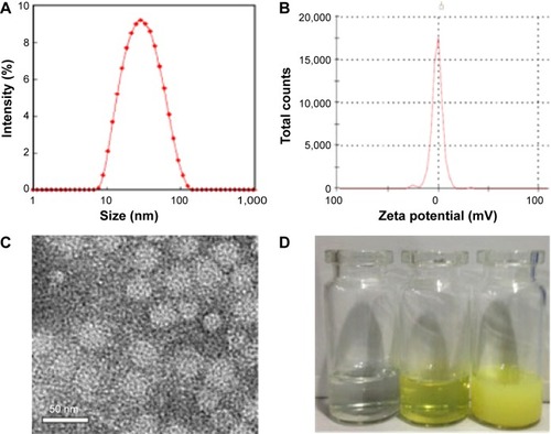
Figure 3 In vitro release study (A) and in vivo pharmacokinetics assay (B) of Qu-M and free-Qu.
Notes: The in vitro release profiles of the Qu-M and free-Qu were examined using a dialysis method, and the in vivo pharmacokinetics was analyzed by high-performance liquid chromatography.
Abbreviations: Qu-M, quercetin-loaded MPEG–PCL nanomicelles; free-Qu, free quercetin; MPEG–PCL, monomethoxy poly(ethylene glycol)–poly(ε-caprolactone).
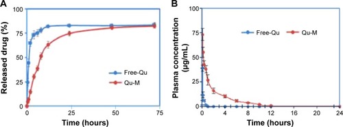
Figure 4 Cytotoxicity of Qu-M and Free-Qu on CT26 cells in vitro.
Notes: Free-Qu as well as Qu-M could efficiently inhibit the viability of C26 cells in a dose-dependent manner after 24 hours (A) and 48 hours (B). Also, Qu-M could enhance the cytotoxic activity of quercetin on C26 cells in vitro.
Abbreviations: Qu-M, quercetin-loaded MPEG–PCL nanomicelles; free-Qu, free quercetin; MPEG–PCL, monomethoxy poly(ethylene glycol)–poly(ε-caprolactone).
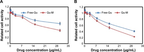
Figure 5 The apoptosis-inducing effects of Qu-M and free-Qu in a dose-dependent manner on CT26 cells in vitro.
Notes: The CT26 cells were incubated with Qu-M, free-Qu, NS, and EM for 48 hours, and Annexin-V- and PI-stained cells were determined by flow cytometry.
Abbreviations: Qu-M, quercetin-loaded MPEG–PCL nanomicelles; free-Qu, free quercetin; NS, normal saline; EM, empty MPEG–PCL nanomicelles; MPEG–PCL, monomethoxy poly(ethylene glycol)–poly(ε-caprolactone); PI, propidium iodide; FITC, fluorescein isothiocyanate.
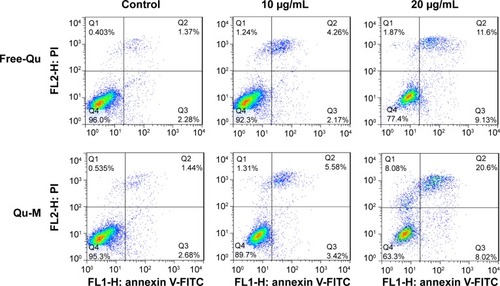
Figure 6 The therapy efficiency of Qu-M and free-Qu on the mice subcutaneous CT26 colon cancer model in vivo.
Notes: Female BALB/c mice were divided into four treatment groups: NS, EM, and free-Qu and Qu-M with a dose of 50 mg/kg. (A) The tumor volumes measured on the indicated days. (B) The body weights of the mice measured on the indicated days. (C) The tumor weights measured on the indicated days. (D) Representative images of tumor gross specimens in each treated group, indicating that Qu-M were more efficacious in repressing the growth of colon tumor in vivo.
Abbreviations: Qu-M, quercetin-loaded MPEG–PCL nanomicelles; free-Qu, free quercetin; NS, normal saline; EM, empty MPEG–PCL nanomicelles; MPEG–PCL, monomethoxy poly(ethylene glycol)–poly(ε-caprolactone).
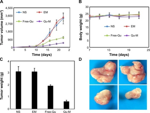
Figure 7 TUNEL assay.
Notes: Tumor tissue sections of NS-treated group (A), EM-treated group (B), free-Qu-treated group (C), and Qu-M-treated group (D) were stained with TUNEL for the cell apoptosis assay. The results indicate that inducing apoptosis may be one of the antitumor mechanisms of Qu-M and free-Qu in vivo.
Abbreviations: NS, normal saline; EM, empty MPEG–PCL nanomicelles; MPEG–PCL, monomethoxy poly(ethylene glycol)–poly(ε-caprolactone); free-Qu, free quercetin; Qu-M, quercetin-loaded MPEG–PCL nanomicelles.
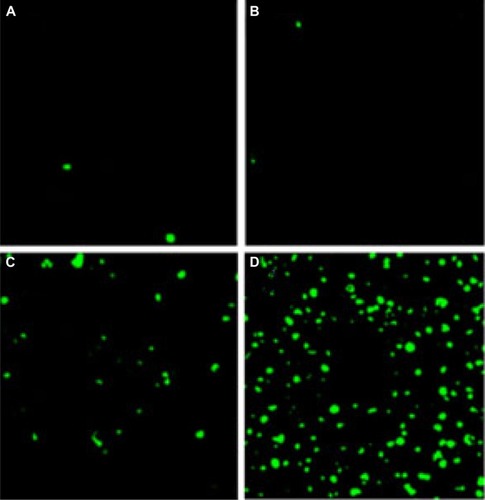
Figure 8 CD31 assay.
Notes: Tumor tissue sections of NS-treated group (A), EM-treated group (B), free-Qu-treated group (C), and Qu-M-treated group (D) were immunostained with CD31 for evaluating the microvessel density. The results imply that antiangiogenesis may be another antitumor mechanism of Qu-M and free-Qu in vivo.
Abbreviations: NS, normal saline; EM, empty MPEG–PCL nanomicelles; MPEG–PCL, monomethoxy poly(ethylene glycol)–poly(ε-caprolactone); free-Qu, free quercetin; Qu-M, quercetin-loaded MPEG–PCL nanomicelles.
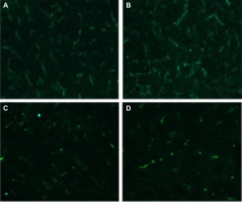
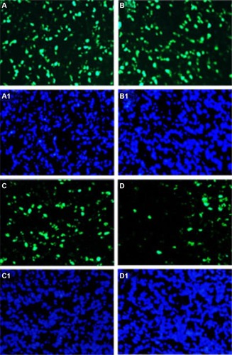
Figure 9 Ki-67 assay.
Notes: Tumor tissue sections of NS-treated group (A, A1), EM-treated group (B, B1), free-Qu-treated group (C, C1), and Qu-M-treated group (D, D1) were immunostained with Ki-67 for evaluating the cell proliferation. The results indicate that antiproliferation may be the other antitumor mechanism of Qu-M and free-Qu in vivo. The green fluorescence indicates the Ki-67 protein (A–D) and the blue fluorescence indicates the total cell nuclei (A1–D1).
Abbreviations: NS, normal saline; EM, empty MPEG–PCL nanomicelles; MPEG–PCL, monomethoxy poly(ethylene glycol)–poly(ε-caprolactone); free-Qu, free quercetin; Qu-M, quercetin-loaded MPEG–PCL nanomicelles.