Figures & data
Figure 1 Preparation of GMP-BSA.
Notes: The hollow porous glucan particles were purified from baker’s yeast, and the cores were synthesized in an electrostatic interaction format. The inner core is the particle formed from BSA and chitosan/tripolyphosphate (TPP)/alginate (Alg), and the outer shell is β-glucan.
Abbreviations: BSA, bovine serum albumin; GMP-BSA, BSA-loaded glucan particles.
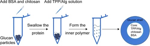
Figure 2 Characterization of GMP-BSA using confocal laser-scanning microscopy, scanning electron microscopy, and dynamic light scattering.
Notes: (A) The fluorescence microscopy images of the GMP-BSA labeled with fluorescein isothiocyanate (FITC; green) or rhodamine B (RhoB; red) on the glucan shells and BSA, respectively. Left, BSA was labeled with FITC, and the glucan shells were labeled with RhoB; right, BSA was labeled with RhoB, and the glucan shells were labeled with FITC. (B) Scanning electron microscopy images of the GMP-BSA (inset shows the GMP-BSA at a higher magnification). (C) Particle size determined by dynamic light scattering.
Abbreviations: BSA, bovine serum albumin; GMP-BSA, BSA-loaded glucan particles; GMP, glucan particles.
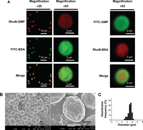
Figure 3 Release profiles of GMP-BSA and protein integrity.
Notes: (A) BSA-release profile of the GMP-BSA in phosphate-buffered saline (pH 7.4) in vitro. (B) Sodium dodecyl sulfate polyacrylamide gel electrophoresis of the released BSA from GMP-BSA stored at 4°C for different times: lane 1, molecular weight markers; lanes 2–4, stored for 5–25 days; lane 5, native BSA. (C) Circular dichroism spectra of the released BSA from the GMP-BSA stored at 4°C for different times.
Abbreviations: BSA, bovine serum albumin; GMP-BSA, BSA-loaded glucan particles.
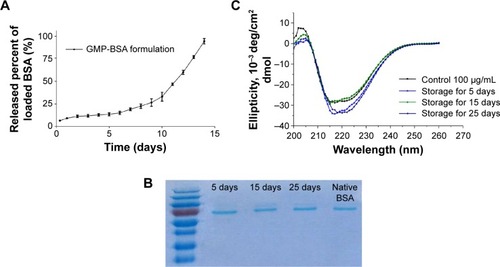
Figure 4 Confocal images of cultured Raw 264.7 cells.
Notes: Cells incubated with different concentrations of GMP-BSA (red/green). GMP-BSA were labeled with fluorescein isothiocyanate (FITC) on BSA (green) and rhodamine B (RhoB) on the glucan shells (red). The nuclei of the cells were stained with 4′,6-diamidino-2-phenylindole (DAPI; blue). Upper panels, ×20 magnification; lower panels, ×63 magnification.
Abbreviations: BSA, bovine serum albumin; GMP-BSA, BSA-loaded glucan particles; GMP, glucan particles.
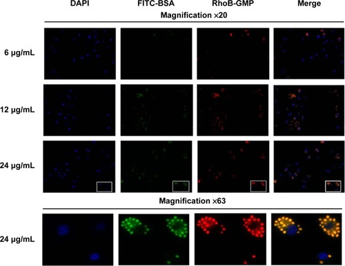
Figure 5 In vitro uptake of GMP-BSA.
Notes: (A) Confocal images of cultured bone marrow-derived macrophage cells incubated with different concentrations of GMP-BSA (red/green). (B) Confocal images of cultured peritoneal exudate macrophages treated with different concentrations of GMP-BSA. The nuclei of the cells were stained with 4′,6-diamidino-2-phenylindole (DAPI; blue), and the membrane was stained with IF488-F4/80 surface antibody (green). The glucan shell of the GMP-BSA was labeled with rhodamine B (RhoB) (red).
Abbreviations: FITC, fluorescein isothiocyanate; GMP-BSA, BSA-loaded glucan particles; BSA, bovine serum albumin; GMP, glucan particles.
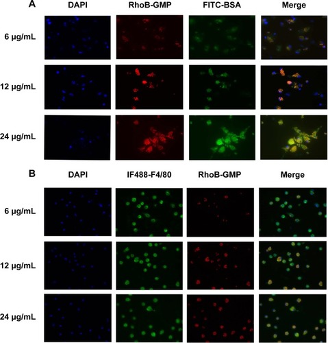
Figure 6 Confocal images.
Notes: Cultured bone marrow-derived macrophages (BMDMs), activated M1 macrophages, or activated M2 macrophages incubated with GMP-BSA for different times (6 hours, 12 hours, or 24 hours). The nuclei of the cells were stained with 4′,6-diamidino-2-phenylindole (blue), and the glucan shell of the GMP-BSA was labeled with rhodamine B (red).
Abbreviations: GMP-BSA, BSA-loaded glucan particles. BSA, bovine serum albumin.
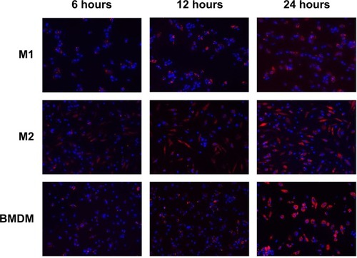
Figure 7 Confocal images.
Notes: Cultured nonphagocytic cells, including NIH3T3, AD293, HeLa, and Caco-2 cells, incubated with GMP-BSA (red/green). The nuclei of the cells were stained with 4′,6-diamidino-2-phenylindole (DAPI; blue). The GMP-BSA were labeled with fluorescein isothiocyanate (FITC) on BSA (green) and rhodamine B (RhoB) on the glucan shells (red).
Abbreviations: GMP-BSA, BSA-loaded glucan particles; BSA, bovine serum albumin; GMP, glucan particles.
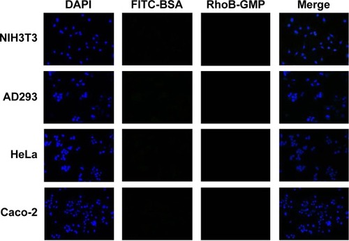
Figure 8 Cytotoxicity of the GMP-BSA to Raw 264.7 cells.
Abbreviations: GMP-BSA, BSA-loaded glucan particles; BSA, bovine serum albumin.
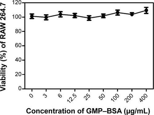
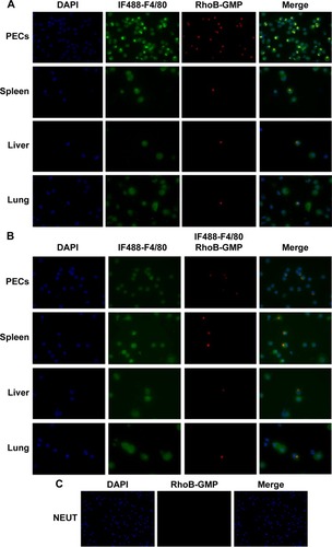
Figure 9 Confocal microscopy images of macrophages.
Notes: Macrophages isolated from the mice intraperitoneally (IP) or intravenously (IV) injected with GMP-BSA, and the neutrophils isolated from the mice IV injected with GMP-BSA. The macrophages in the peritoneal exudate cells (PECs) and organs and the neutrophils in the blood from the IV injected mice were isolated for microscopic observation. (A) Macrophages from the IP injected mice. (B) Macrophages from the IV injected mice. (C) Neutrophils (NEUT) from the IV injected mice. The GMP-BSA were labeled with rhodamine B (RhoB; red) (red) on the glucan shells. The nuclei of the cells were stained with 4′,6-diamidino-2-phenylindole (DAPI; blue), and the membrane of the macrophages was stained with IF488-F4/80 surface antibody (green).
Abbreviations: GMP-BSA, BSA-loaded glucan particles; BSA, bovine serum albumin; GMP, glucan particles.