Figures & data
Figure 1 Histogram showing the particle size and size distribution of INPs.
Abbreviation: INPs, imatinib mesylate-loaded poly(lactide-co-glycolide) nanoparticles.
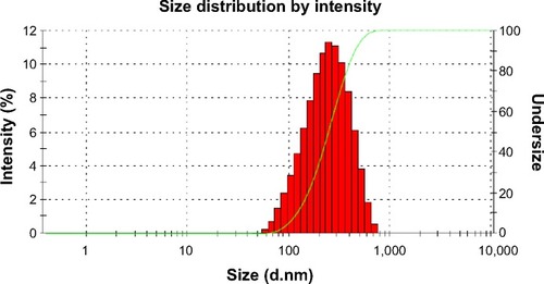
Figure 2 Scanning electron microscopic image of INPs (scale bar =2 μm).
Abbreviation: INPs, imatinib mesylate-loaded poly(lactide-co-glycolide) nanoparticles.
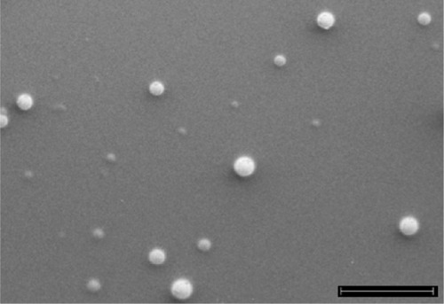
Figure 3 In vitro drug release characteristics of INPs, showing a biphasic profile, an initial burst release up to 24 hours, and then an extended release over 10 days.
Abbreviations: INPs, imatinib mesylate-loaded poly(lactide-co-glycolide) nanoparticles.
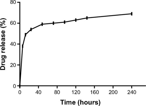
Figure 4 Internalization of CNPs.
Notes: (A) Red fluorescence from LysoTracker® Red showing lysosomes and acidic vesicles; (B) green fluorescence from curcumin showing intracellular localization of CNPs; (C) overlap image of both figures (A and B) resulted in yellow spots showing that CNPs are in the lysosomes, and acidic vesicles of mature endosomes.
Abbreviation: CNPs, curcumin-loaded poly(lactide-co-glycolide) nanoparticles.
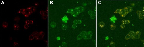
Figure 5 MTT assay results showing higher cytotoxicity of INPs compared to free imatinib mesylate. Imatinib concentration of 0.0 μM represents respective controls of INPs and free drug, which showed 100% cell viability.
Abbreviations: INPs, imatinib mesylate-loaded poly(lactide-co-glycolide) nanoparticles; MTT, methylthiazolyldiphenyl-tetrazolium bromide.
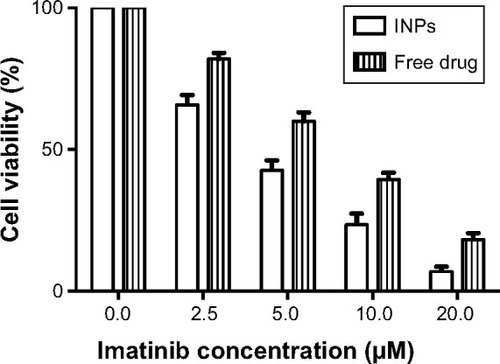
Table 1 Subacute in vivo toxicity analyses of rats after 28 days
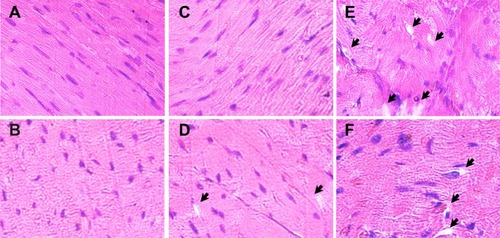
Figure 6 Histopathology of myocardium from experimental animals. Representative images of left ventricular histology from experimental animals stained with hematoxylin and eosin are shown.
Notes: (A, B) The cardiac myofibrils from control rats exhibited regular arrangement of thick and thin myofilaments. (C, D) INP treatment did not alter the morphology of myocardium of rats significantly, although very few isolated vacuoles are present. (E, F) Myofibrillar loss and formation of variable-sized cytoplasmic vacuoles in the myocardium of rats treated with 50 mg/kg dose of free imatinib mesylate. Vacuole structures are indicated with black arrows.
Abbreviation: INP, imatinib mesylate-loaded poly(lactide-co-glycolide) nanoparticle.