Figures & data
Figure 1 Scanning electron micrographs of PLLAsc (A) and APLLAsc (B).
Notes: A drop of PLLAsc and APLLAsc suspensions in isopropanol was deposited on the sample holder. The magnitude scale bars are indicated.
Abbreviations: PLLAsc, poly(l-lactide) single crystals; APLLAsc, amino-functionalized poly(l-lactide) single crystals.
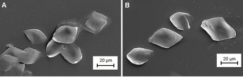
Figure 2 Attenuated total reflection Fourier transform infrared spectra of PLLAsc and APLLAsc (A). Differential scanning calorimetry thermograms of PLLAsc and APLLAsc recorded at 10°C per minute (B).
Note: The arrow indicates the amide C=O stretching and N-Hbending large peak (A).
Abbreviations: PLLAsc, poly(l-lactide) single crystals; APLLAsc, amino-functionalized poly(l-lactide) single crystals.
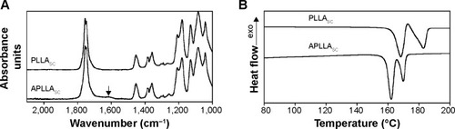
Table 1 Advancing and receding contact angles of pristine and APLLA films in water measured using a stage speed of 60 μm per second
Figure 3 Protein release experiment.
Notes: E7-PLLAsc and E7-APLLAsc suspensions were centrifuged and rinsed in phosphate-buffered saline five times. The released E7 was quantify in the supernatant after each rinse step. The y axis reports the cumulative quantity of E7 released with respect to that initially adsorbed on the PLLA substrates. The x axis indicates each single wash step. The values are the mean of three determinations.
Abbreviations: PLLA, poly(l-lactide); E7-PLLAsc, E7-containing poly(l-lactide) single crystals; E7-APLLAsc, E7-containing amino-functionalized poly(l-lactide) single crystals.
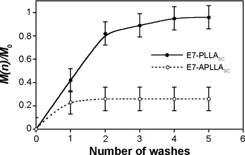
Figure 4 Scanning electron micrographs of E7-PLLAsc (A) and E7-APLLAsc (B) aggregates.
Notes: A drop of either E7-PLLAsc or E7-APLLAsc suspension, with the same concentration employed for subcutaneous mouse immunization, was deposited on the sample holder. The magnitude scale bars are indicated.
Abbreviations: E7-PLLAsc, E7-containing poly(l-lactide) single crystals; E7-APLLAsc, E7-containing amino-functionalized poly(l-lactide) single crystals.
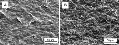
Table 2 Sera pool titration by endpoint dilution ELISA of antibodies against E7 after the second and third doses of vaccine
Figure 5 Determination of immunoglobulin isotypes by enzyme-linked immunosorbent assay.
Notes: The E7-specific IgG1, IgG2b, IgG2c and IgG3 immune reactivity is shown in OD450 values on the y axis. The bars represent pools from sera of mice groups immunized with free E7 (E7, white bars), E7-PLLAsc (gray bars), and E7-APLLAsc (black bars). The four isotypes are indicated on the x axis along with the result of a total IgG enzyme-linked immunosorbent assay performed in parallel as control using the same serum pools. *Statistically significant result (P<0.05).
Abbreviations: E7-PLLAsc, E7-containing poly(l-lactide) single crystals; E7-APLLAsc, E7-containing amino-functionalized poly(l-lactide) single crystals; Ig, immun oglobulin.
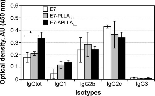
Figure 6 INF-γ-secreting cells from mice immunized with E7-APLLAsc, E7-PLLAsc and free E7 (E7).
Notes: The cells were stimulated with either an unrelated mixture of peptides (unp, black bars) or two CTLs E7 peptides (pE7, grey bars). *P=0.009; **P=0.01.
Abbreviations: unp, unrelated mixture of peptides; pE7, E7 peptide; IFN-γ, interferon gamma; E7-PLLAsc, E7-containing poly(l-lactide) single crystals; E7-APLLAsc, E7-containing amino-functionalized poly(l-lactide) single crystals.
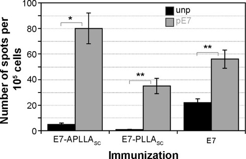
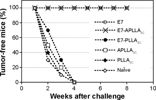
Figure 7 Tumor protection experiment.
Notes: Mice naïve or vaccinated with three doses of free E7 (E7), E7-APLLAsc, E7-PLLAsc, APLLAsc and PLLAsc were challenged with 1×10⁵ TC-1 tumor cells and tumor growth was monitored weekly. The x axis indicates weeks of monitoring after tumor challenge and the y axis indicates the percentages of animals without tumor.
Abbreviations: E7-PLLAsc, E7-containing poly(l-lactide) single crystals; E7-APLLAsc, E7-containing amino-functionalized poly(l-lactide) single crystals.