Figures & data
Figure 1 Transmission electron microscopy photomicrograph (magnification 50,000×) (A) and particle size distribution (B) of the pFDNA-SiO2-NPs.
Abbreviation: pFDNA-SiO2-NPs, Newcastle disease virus F gene encapsulated in the SiO2 nanoparticles.
Figure 2 Detection of SiO2 nanoparticles loaded with plasmid DNA for connection, release, and protection properties.
Notes: (A) Agarose gel electrophoresis of the amino-modified silica-DNA complexes. Maker (M): DL 15,000 marker. Lane 1: plasmid DNA. Lanes 2–8: the mass ratios of SiO2 to plasmid DNA were 1:5, 1:1, 5:1, 10:1, 20:1, 30:1, and 40:1, respectively. (B) Stability analysis of the plasmid DNA binding with SiO2 nanoparticles. (C) Analysis of protection of the plasmid DNA from DNase I digestion. Maker (M): DL 15,000 marker. Lane 1: plasmid DNA. Lanes 2–8: the mass ratios of SiO2 to plasmid DNA were 1:5, 1:1, 5:1, 10:1, 20:1, 30:1, and 40:1, respectively.
Figure 3 Transmission electron photomicrographs of the pFDNA-SiO2-NPs (A), LDH (B), and pFDNA-LDH@SiO2-NPs (C–E). Note: Magnification: 100,000×.
Abbreviations: LDH, layered double hydroxide; NPs, nanoparticles; pFDNA-LDH@SiO2-NPs, Newcastle disease virus F gene encapsulated in LDH@SiO2-NPs; pFDNA-SiO2-NPs, Newcastle disease virus F gene encapsulated in the SiO2 nanoparticles.
Figure 4 Size distribution of LDH (A) and pFDNA-LDH@SiO2-NPs (B).
Notes: Measurement of these particles showed a narrow distribution pattern of LDH and pFDNA-LDH@SiO2-NPs, and their average diameters were 99.1 nm and 371.93 nm, respectively.
Abbreviations: LDH, layered double hydroxide; NPs, nanoparticles; pFDNA-LDH@SiO2-NPs, Newcastle disease virus F gene encapsulated in LDH@SiO2-NPs.
Figure 5 Fourier transform infrared spectroscopy spectrum (A) and XRD curve (B) for each of the LDH and pFDNA-LDH@SiO2-NPs.
Notes: The XRD patterns of the pFDNA-LDH@SiO2-NPs exhibited typical diffractions of LDH including (003) and (006), indicative of the presence of LDH crystallite. The characteristic peak at 20° of pFDNA-LDH@SiO2-NPs showed the existence of SiO2.
Abbreviations: LDH, layered double hydroxide; NPs, nanoparticles; pFDNA-LDH@SiO2-NPs, Newcastle disease virus F gene encapsulated in LDH@SiO2-NPs; XRD, X-ray diffraction.
Figure 6 In vitro release profiles of DNA from pFDNA-LDH@SiO2-NPs in phosphate-buffered saline (pH 7.4).
Notes: The experiment was repeated three times and measured in triplicate. Data are presented as the mean ± standard deviation (n=3).
Abbreviations: LDH, layered double hydroxide; NPs, nanoparticles; pFDNA-LDH@SiO2-NPs, Newcastle disease virus F gene encapsulated in LDH@SiO2-NPs.
Figure 7 In vitro expression of the pFDNA-LDH@SiO2-NPs in 293 T cells assayed by indirect immunofluorescence (×40).
Notes: (A) Naked plasmid pVAX1-F(o) DNA group. (B) Transfected pFDNA-LDH@SiO2-NPs group. (C) Blank LDH@SiO2-NPs group. (D) 293 T cell group as the negative control.
Abbreviations: LDH, layered double hydroxide; NPs, nanoparticles; pFDNA-LDH@SiO2-NPs, Newcastle disease virus F gene encapsulated in LDH@SiO2-NPs.
Figure 8 Histopathological analyses of normal duodena, proventriculi, and myocardia.
Notes: (A) Tissues of duodena were immunized with pFDNA-LDH@SiO2-NPs IM (a), pFDNA-LDH@SiO2-NPs IN (b), and PBS IM (c). (B) Tissues of proventriculi were immunized with pFDNA-LDH@SiO2-NPs IM (a), pFDNA-LDH@SiO2-NPs IN (b), and PBS IM (c). (C) Tissues of myocardia were immunized with pFDNA-LDH@SiO2-NPs IM (a), pFDNA-LDH@SiO2-NPs IN (b), and PBS IM (c).
Abbreviations: IM, intramuscularly; IN, intranasally; LDH, layered double hydroxide; NPs, nanoparticles; PBS, phosphate-buffered saline; pFDNA-LDH@SiO2-NPs, Newcastle disease virus F gene encapsulated in LDH@SiO2-NPs.
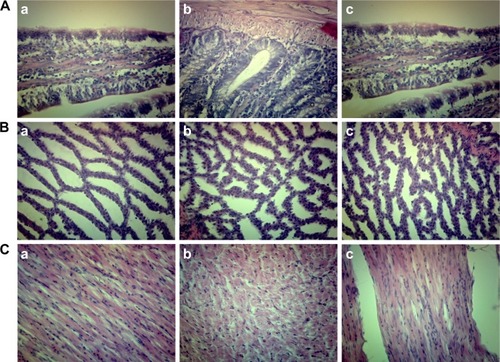
Table 1 IgG antibody titers in serum samples of specific pathogen-free chickens immunized with PBS IM, LDH@SiO2-NPs IM, naked plasmid pVAX1-F(o) DNA IM, pFDNA-LDH@ SiO2-NPs IM, and pFDNA-LDH@SiO2-NPs IN (unit: log 2), respectively
Figure 9 IgA antibody levels in serum (A), tracheal fluid (B), bile (C), and Harderian glands (D) of specific pathogen-free chickens immunized with PBS IM, blank LDH@SiO2-NPs IM, naked plasmid pVAX1-F(o) DNA IM, pFDNA-LDH@SiO2-NPs IM, and pFDNA-LDH@SiO2-NPs IN.
Notes: IgA antibody levels are presented as mean ± standard deviation of five experiments. Data with different small letters show significant difference (P<0.05).
Abbreviations: IM, intramuscularly; IN, intranasally; LDH, layered double hydroxide; NPs, nanoparticles; PBS, phosphate-buffered saline; pFDNA-LDH@SiO2-NPs, Newcastle disease virus F gene encapsulated in the LDH@SiO2-NPs.
Table 2 The stimulation index of T lymphocyte proliferation in specific pathogen-free chickens post-immunization
Table 3 Protection of the immunized specific pathogen-free chickens after being challenged with the highly virulent Newcastle disease virus strain F48E9
Figure 10 Histopathological analyses of the duodena and myocardia of chickens challenged with the highly virulent NDV strain F48E9.
Notes: (A) Tissues of the duodena from chickens immunized with pFDNA-LDH@SiO2-NPs IN. (B) Tissues of the myocardia from chickens immunized with pFDNA-LDH@SiO2-NPs IN. (C) Tissues of the duodena from chickens immunized with pFDNA-LDH@SiO2-NPs IM, naked plasmid pVAX1-F(o) DNA IM, or PBS IM. (D) Tissues of the myocardia from chickens in groups immunized with the pFDNA-LDH@SiO2-NPs IM, naked plasmid pVAX1-F(o) DNA IM, or PBS IM.
Abbreviations: IM, intramuscularly; IN, intranasally; LDH, layered double hydroxide; NDV, Newcastle disease virus; NPs, nanoparticles; PBS, phosphate-buffered saline; pFDNA-LDH@SiO2-NPs, NDV F gene encapsulated in the LDH@SiO2-NPs.
Figure S1 Preparation of pFDNA-LDH@SiO2-NPs.
Abbreviations: APTES, 3-aminopropyl-triethoxysilane; LDH, layered double hydroxide; NPs, nanoparticles; pFDNA-LDH@SiO2-NPs, Newcastle disease virus F gene encapsulated in LDH@SiO2-NPs; pFDNA-SiO2-NPs, Newcastle disease virus F gene encapsulated in SiO2-NPs; TEOS, tetraethyl orthosilicate.