Figures & data
Figure 1 (A) Experimental set-up, (B) temperature curves, and (C) X-ray image during magnetic thermoablation.
Notes: (A) Scheme of the used experimental set-up. Nanoparticles are indicated by orange spots. (B) Representative temperature curves of different tumor regions during treatment. Body temperature was observed by thermal measurement via the rectum. Higher temperatures in the central tumor region correlate with the distribution of the administered magnetic material. After reaching ablative temperatures of approximately 65°C, the AMF was shut-off. (C) X-ray images of mice were used to control MNP relocalization and the localization of thermocouples for thermal treatment.
Abbreviations: AMF, alternating magnetic field; MNP, magnetic nanoparticles.
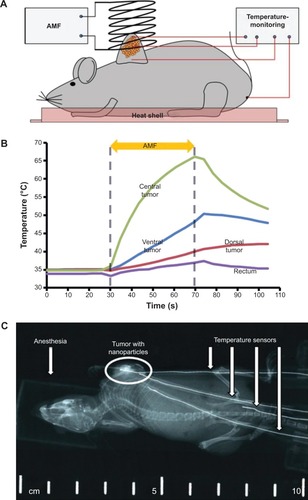
Figure 2 Effect of magnetic thermoablation on the BCL2, HSP70, and FGF-R1 protein expression pattern in BT474 tumors.
Notes: Representative pictures after IHC staining of BCL2, HSP70, and FGF-R1 were chosen. Blue: nuclei, red: protein-specific antibody, scale bar: 100 μm. Relative semi-quantitative expression of investigated proteins is depicted in diagrams. In diagrams, bars represent the relative number of slides with a specific protein expression normalized to the total number of all slides in the specific treatment group. Bar colors represent the amount of specifically stained area within the viable tumor region: unstained/negative (light gray), less than 50% of vital area stained (dark grey), more than 50% of vital area stained (black). Whereas a downregulation of BCL2 and FGF-R1 can be assumed, HSP70 expression remained unchanged.
Abbreviations: BCL2, B-cell lymphoma 2; HSP70, heat shock protein; FGF-R1, fibroblast growth factor receptor 1; IHC, immunohistochemistry; MNP, magnetic nanoparticles; AMF, alternating magnetic field.
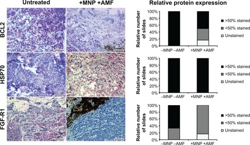
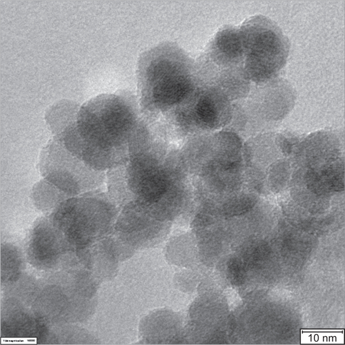
Figure S1 High-resolution transmission electron microscopy (HRTEM) micrograph of 200 nm fluidMAG-D magnetic nanoparticles (MNP).
Notes: HRTEM micrograph reveals smaller core particles of approximately 10–12 nm in size clustered to a particle core size of approximately 70 nm in total. The HRTEM micrograph was prepared as described elsewhere.Citation37