
Figures & data
Table 1 Primer sequences
Figure 1 Synthesis of five copies of M2e coding region with PCR.
Notes: (A) Primer-overlapping PCR for generating the coding sequence of five copies of M2e. The overlapping of fragments 1, 2, 3, and 4 eventually gives rise to a full-length M2ex5 through multiple cycles of PCR reaction. (B) Nucleotide and amino acid sequences of the M2ex5 coding region and polypeptide, respectively. Three glycine residues (boxed) were added in between each M2e gene, and also at both terminal ends of the M2ex5 gene as linkers. Underlined nucleotide sequences indicate overlapping region of primers used for gene synthesis. The nucleotide sequence was modified to have different sequences encoding for the same amino acids.
Abbreviations: PCR, polymerase chain reaction; M2ex5, nucleotide sequence encoding five copies of matrix 2 ectodomain of influenza A virus; M2e, influenza A matrix 2 ectodomain.
Figure 2 Primary structures and amino acid sequences of NvC, NvC-M2ex1, NvC-M2ex3, and NvC-M2ex5.
Notes: One, three, and five copies of M2e were fused to the C-terminal end of the MrNV capsid protein. Horizontal lines indicate the absence of amino acid sequence. The amino acid sequences of MrNV capsid protein, linker, M2e, myc epitope, and His-tag are shown.
Abbreviations: MrNV, Macrobrachium rosenbergii nodavirus; NvC, Macrobrachium rosenbergii nodavirus capsid protein; NvC-M2ex1, Macrobrachium rosenbergii nodavirus capsid protein displaying one copy of influenza A matrix 2 ectodomain; NvC-M2ex3, Macrobrachium rosenbergii nodavirus capsid protein displaying three copies of influenza A matrix 2 ectodomain; NvC-M2ex5, Macrobrachium rosenbergii nodavirus capsid protein displaying five copies of influenza A matrix 2 ectodomain; M2e, influenza A matrix 2 ectodomain.
Figure 3 Expression and purification of the recombinant proteins.
Notes: Total cell lysate proteins were analyzed on (A) SDS-PAGE, and (B) Western blotting. “−” refers to non-induced cell lysate samples, whereas “+” refers to IPTG-induced cell lysate samples from Escherichia coli expressing the NvC-M2ex1, NvC-M2ex3, and NvC-M2ex5. On both SDS-PAGE and Western blot, protein bands of approximately 50 kDa, 56 kDa, and 63 kDa can be observed upon induction (indicated by arrow heads). Purified NvC-M2ex1, NvC-M2ex3, and NvC-M2ex5 proteins were analyzed with (C) SDS-PAGE and (D) Western blotting.
Abbreviations: SDS-PAGE, sodium dodecyl sulfate polyacrylamide gel electrophoresis; IPTG, isopropyl β-D-1-thiogalactopyranoside; NvC-M2ex1, Macrobrachium rosenbergii nodavirus capsid protein displaying one copy of influenza A matrix 2 ectodomain; NvC-M2ex3, Macrobrachium rosenbergii nodavirus capsid protein displaying three copies of influenza A matrix 2 ectodomain; NvC-M2ex5, Macrobrachium rosenbergii nodavirus capsid protein displaying five copies of influenza A matrix 2 ectodomain; M, protein markers with their corresponding molecular masses in kDa.
Figure 4 Transmission electron microscopic analysis of fusion proteins.
Notes: (A) NvC-M2ex1, (B) NvC-M2ex3, and (C) NvC-M2ex5 were negatively stained with uranyl acetate and the micrographs were taken under 100,000× magnification. Spherical VLPs of approximately 30 nm in diameter were observed for all the three fusion proteins.
Abbreviations: NvC-M2ex1, Macrobrachium rosenbergii nodavirus capsid protein displaying one copy of influenza A matrix 2 ectodomain; NvC-M2ex3, Macrobrachium rosenbergii nodavirus capsid protein displaying three copies of influenza A matrix 2 ectodomain; NvC-M2ex5, Macrobrachium rosenbergii nodavirus capsid protein displaying five copies of influenza A matrix 2 ectodomain; VLPs, virus-like particles.
Figure 5 Antigenicity of M2e displayed on NvC.
Notes: Microtiter plate wells were coated with different concentrations of fusion proteins and the synthetic M2e peptide. The wells were blocked with milk diluents and the anti-influenza A M2 antibody was added to react with the coated proteins. NvC-M2ex1, NvC-M2ex3, and NvC-M2ex5 were compared with the synthetic M2e peptide (positive control) and the NvC (negative control).
Abbreviations: M2e, influenza A matrix 2 ectodomain; M2, influenza A matrix 2 protein; NvC, Macrobrachium rosenbergii nodavirus capsid protein; NvC-M2ex1, Macrobrachium rosenbergii nodavirus capsid protein displaying one copy of influenza A matrix 2 ectodomain; NvC-M2ex3, Macrobrachium rosenbergii nodavirus capsid protein displaying three copies of influenza A matrix 2 ectodomain; NvC-M2ex5, Macrobrachium rosenbergii nodavirus capsid protein displaying five copies of influenza A matrix 2 ectodomain.
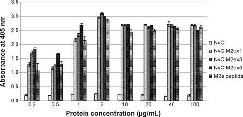
Figure 6 Immunogenicity of the fusion proteins in BALB/c mice.
Notes: Antibodies specifically against M2e in serum samples collected at week 2 (pre-injection), week 5 (3 weeks after first injection), week 8 (3 weeks after first booster injection), and week 9 (1 week after second booster injection) were measured by ELISA. Serum samples from mice were pooled according to the groups injected with HEPES buffer, NvC, NvC-M2ex1, NvC-M2ex3, and NvC-M2ex5. The serum samples were then reacted with synthetic M2e peptide coated onto microtiter plate wells. Alphabets and roman numerals (a, b, c, d; x, y, z; and I, II, III, IV) on top of each bar (group) represent statistical significance (P<0.001) of results within each time point. The asterisks (**) on top of the bar chart show extreme significance (P<0.0001) compared with the group of mice immunized with NvC (control without M2e).
Abbreviations: M2e, influenza A matrix 2 ectodomain; ELISA, enzyme-linked immunosorbent assay; HEPES, 2-[4-(2-hydroxyethyl)piperazin-1-yl]ethanesulfonate; NvC, Macrobrachium rosenbergii nodavirus capsid protein; NvC-M2ex1, Macrobrachium rosenbergii nodavirus capsid protein displaying one copy of influenza A matrix 2 ectodomain; NvC-M2ex3, Macrobrachium rosenbergii nodavirus capsid protein displaying three copies of influenza A matrix 2 ectodomain; NvC-M2ex5, Macrobrachium rosenbergii nodavirus capsid protein displaying five copies of influenza A matrix 2 ectodomain.
![Figure 6 Immunogenicity of the fusion proteins in BALB/c mice.Notes: Antibodies specifically against M2e in serum samples collected at week 2 (pre-injection), week 5 (3 weeks after first injection), week 8 (3 weeks after first booster injection), and week 9 (1 week after second booster injection) were measured by ELISA. Serum samples from mice were pooled according to the groups injected with HEPES buffer, NvC, NvC-M2ex1, NvC-M2ex3, and NvC-M2ex5. The serum samples were then reacted with synthetic M2e peptide coated onto microtiter plate wells. Alphabets and roman numerals (a, b, c, d; x, y, z; and I, II, III, IV) on top of each bar (group) represent statistical significance (P<0.001) of results within each time point. The asterisks (**) on top of the bar chart show extreme significance (P<0.0001) compared with the group of mice immunized with NvC (control without M2e).Abbreviations: M2e, influenza A matrix 2 ectodomain; ELISA, enzyme-linked immunosorbent assay; HEPES, 2-[4-(2-hydroxyethyl)piperazin-1-yl]ethanesulfonate; NvC, Macrobrachium rosenbergii nodavirus capsid protein; NvC-M2ex1, Macrobrachium rosenbergii nodavirus capsid protein displaying one copy of influenza A matrix 2 ectodomain; NvC-M2ex3, Macrobrachium rosenbergii nodavirus capsid protein displaying three copies of influenza A matrix 2 ectodomain; NvC-M2ex5, Macrobrachium rosenbergii nodavirus capsid protein displaying five copies of influenza A matrix 2 ectodomain.](/cms/asset/12ab8666-8a3c-46a7-b22d-0d148cce6eda/dijn_a_77405_f0006_b.jpg)
Figure 7 Immunophenotyping of mice splenocytes.
Notes: Spleens from mice immunized with HEPES, NvC, NvC-M2ex1, NvC-M2ex3, and NvC-M2ex5 were harvested at week 9. The spleens were meshed through cell strainers to obtain single cell suspensions. The splenocytes were then probed with two sets of fluorochrome-labeled antibodies for detection of the (A) CD8+:CD4+ population ratio and (B) F4/80+ population. Roman numerals and alphabets (I, II; x, y, z) on top of each bar (group) represent the statistical significance (P<0.05) of results between different treatment groups.
Abbreviations: HEPES, 2-[4-(2-hydroxyethyl)piperazin-1-yl]ethanesulfonate; NvC, Macrobrachium rosenbergii nodavirus capsid protein; NvC-M2ex1, Macrobrachium rosenbergii nodavirus capsid protein displaying one copy of influenza A matrix 2 ectodomain; NvC-M2ex3, Macrobrachium rosenbergii nodavirus capsid protein displaying three copies of influenza A matrix 2 ectodomain; NvC-M2ex5, Macrobrachium rosenbergii nodavirus capsid protein displaying five copies of influenza A matrix 2 ectodomain; CD8+, cells possessing clusters of differentiation 8 (cytotoxic T lymphocytes); CD4+, cells possessing clusters of differentiation 4 (T helper cells); F4/80+, cells possessing F4/80 cell surface marker (macrophages).
![Figure 7 Immunophenotyping of mice splenocytes.Notes: Spleens from mice immunized with HEPES, NvC, NvC-M2ex1, NvC-M2ex3, and NvC-M2ex5 were harvested at week 9. The spleens were meshed through cell strainers to obtain single cell suspensions. The splenocytes were then probed with two sets of fluorochrome-labeled antibodies for detection of the (A) CD8+:CD4+ population ratio and (B) F4/80+ population. Roman numerals and alphabets (I, II; x, y, z) on top of each bar (group) represent the statistical significance (P<0.05) of results between different treatment groups.Abbreviations: HEPES, 2-[4-(2-hydroxyethyl)piperazin-1-yl]ethanesulfonate; NvC, Macrobrachium rosenbergii nodavirus capsid protein; NvC-M2ex1, Macrobrachium rosenbergii nodavirus capsid protein displaying one copy of influenza A matrix 2 ectodomain; NvC-M2ex3, Macrobrachium rosenbergii nodavirus capsid protein displaying three copies of influenza A matrix 2 ectodomain; NvC-M2ex5, Macrobrachium rosenbergii nodavirus capsid protein displaying five copies of influenza A matrix 2 ectodomain; CD8+, cells possessing clusters of differentiation 8 (cytotoxic T lymphocytes); CD4+, cells possessing clusters of differentiation 4 (T helper cells); F4/80+, cells possessing F4/80 cell surface marker (macrophages).](/cms/asset/9c31c979-f649-4218-8cbb-1ce97f353945/dijn_a_77405_f0007_b.jpg)
Table 2 Cytokine concentrations in pooled serum samples