Figures & data
Figure 1 (A) The formation of PTX-loaded polymeric nanoparticles from PCL-PEG-PCL. (B) TEM image of PTX-loaded PCEP NPs.
Notes: Left scale bar =500 nm, right scale bar =200 nm.
Abbreviations: TEM, transmission electron microscopy; PCL-PEG-PCL, poly(ε-caprolactone)–poly(ethylene glycol)–poly(ε-caprolactone); PTX, paclitaxel; PCEP NPs, polymeric nanoparticles formed from PCL-PEG-PCL; PEG, poly(ethylene glycol); PCL, poly(ε-caprolactone).
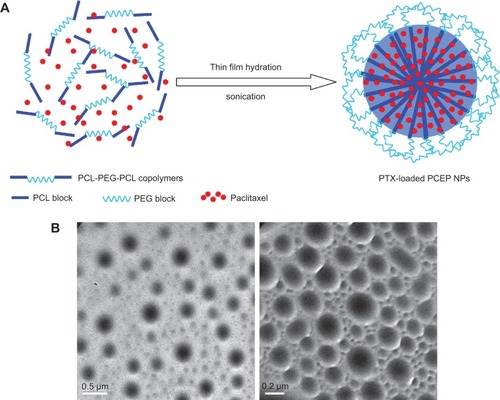
Figure 2 Characterization of PTX-loaded folate targeted nanoparticles of mixed lipid-shell and polymer core (FLPNPs).
Notes: (A) Schematic illustration of the formulation of PTX-loaded FLPNPs. (B) TEM image of PTX-loaded FLPNPs (left scale bar =200 nm, right scale bar =100 nm). (C) Confocal laser scanning fluorescence image of rhodamine-PE labeled PTX-loaded FLPNPs (scale bar =2 μm): (a) Rhodamine channel, (b) Transmitted light, (c) Merged.
Abbreviations: FLPNPs, folate modified lipid-shell and polymer-core nanoparticles; PCL-PEG-PCL, poly(ε-caprolactone)–poly(ethylene glycol)–poly(ε-caprolactone); DSPE-PEG2000, 1,2-distearoyl-sn-glycero-3-phosphoethanolamine-N-[methoxy (polyethylene glycol)-2000]; PTX, paclitaxel; TEM, transmission electron microscopy.
![Figure 2 Characterization of PTX-loaded folate targeted nanoparticles of mixed lipid-shell and polymer core (FLPNPs).Notes: (A) Schematic illustration of the formulation of PTX-loaded FLPNPs. (B) TEM image of PTX-loaded FLPNPs (left scale bar =200 nm, right scale bar =100 nm). (C) Confocal laser scanning fluorescence image of rhodamine-PE labeled PTX-loaded FLPNPs (scale bar =2 μm): (a) Rhodamine channel, (b) Transmitted light, (c) Merged.Abbreviations: FLPNPs, folate modified lipid-shell and polymer-core nanoparticles; PCL-PEG-PCL, poly(ε-caprolactone)–poly(ethylene glycol)–poly(ε-caprolactone); DSPE-PEG2000, 1,2-distearoyl-sn-glycero-3-phosphoethanolamine-N-[methoxy (polyethylene glycol)-2000]; PTX, paclitaxel; TEM, transmission electron microscopy.](/cms/asset/dd46dd31-b890-4128-a6cf-d36a0bf62a98/dijn_a_77667_f0002_c.jpg)
Table 1 Physicochemical characterization of PTX-loaded PCEP NPs, LPNPs, and FLPNPs
Figure 3 In vitro PTX release profiles from LPNPs and FLPNPs in 1.0 M sodium salicylate solutions (n=3).
Abbreviations: PTX, paclitaxel; LPNPs, lipid–polymer hybrid nanoparticles; FLPNPs, folate modified lipid-shell and polymer-core nanoparticles.
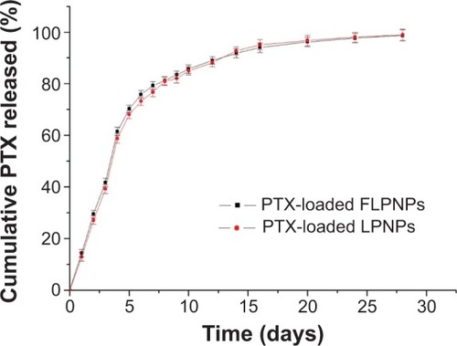
Figure 4 CLSM images of FR-positive EMT6 cells and FR-negative L929 cells after incubation with Nile Red-loaded LPNPs and FLPNPs for 2 h.
Notes: Row 1 and 2: EMT6 cells were used. Row 3 and 4: L929 cells were used. In row 1 and 3, LPNPs were used while in row 2 and 4, FLPNPs were used.
Abbreviations: FITC, fluorescein isothiocyanate; CLSM, confocal laser scanning microscopy; FR, folate receptor; LPNPs, lipid–polymer hybrid nanoparticles; FLPNPs, folate modified lipid-shell and polymer-core nanoparticles; h, hours.
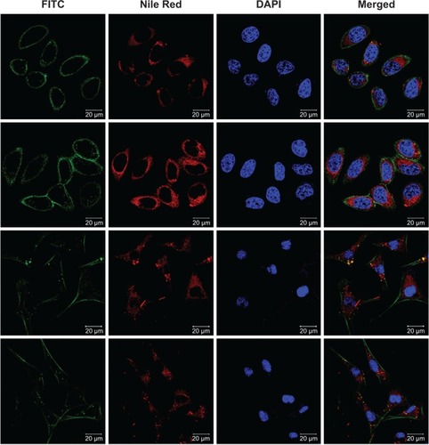
Figure 5 The in vitro cellular uptake efficiency of EMT6 cells after incubation with Nile Red-loaded LPNPs and FLPNPs for 0.5, 2, and 24 h (n=6). *P<0.05.
Abbreviations: PTX, paclitaxel; LPNPs, lipid–polymer hybrid nanoparticles; FLPNPs, folate modified lipid-shell and polymer-core nanoparticles; h, hours.
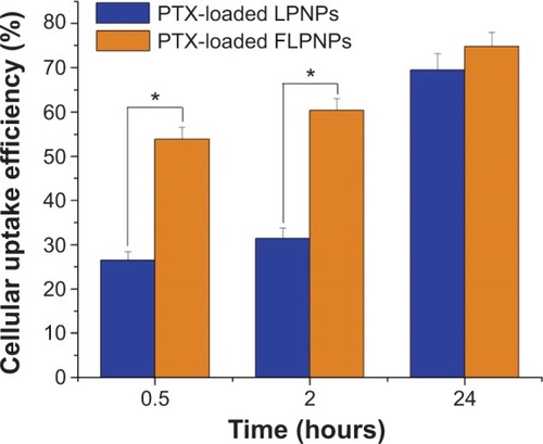
Figure 6 The in vitro cell viability of EMT6 cells at various concentrations of the drug under 24, 48, and 72 h treatment with Taxol, PTX-loaded LPNPs, and PTX-loaded FLPNPs (n=6). *P<0.05.
Abbreviations: PTX, paclitaxel; LPNPs, lipid–polymer hybrid nanoparticles; FLPNPs, folate modified lipid-shell and polymer-core nanoparticles; h, hours.
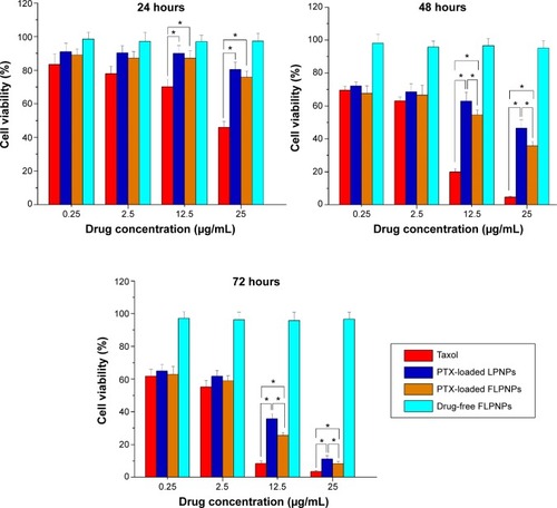
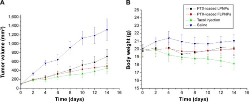
Figure 7 In vivo antitumor efficacy (A) and body weight changes (B) after intratumoral injection of PTX-loaded LPNPs, PTX-loaded FLPNPs, Taxol injection, and saline on EMT6 tumor-bearing BALB/c mice. Each point represents the mean of tumor size ± SEM (n=10).
Abbreviations: PTX, paclitaxel; LPNPs, lipid–polymer hybrid nanoparticles; FLPNPs, folate modified lipid-shell and polymer-core nanoparticles; SEM, standard error of the mean.