Figures & data
Table 1 Physicochemical characteristics of DEX-NPs
Figure 1 Outline of animal experiments.
Notes: There were seven animal experiments – namely, the in vivo distribution of Coumarin 6 NPs (n=6), the in vivo release of DEX from DEX-NPs (N=35), the hearing assessment of the sham operation (N=6), the establishment of a hearing loss animal model (N=18), the protection of hearing function by DEX-NPs – ABR (N=52), the protection of hearing function by DEX-NPs – hair cell counting (N=24), and inflammatory response to NPs (N=6).
Abbreviations: NP, nanoparticle; n, number; DEX, dexamethasone; DEX-NPs, dexamethasone-loaded polyethylene glycol-coated polylactic acid stealth nanoparticles; ABR, auditory brainstem response; CDDP, cis-diamminedichloroplatinumII.
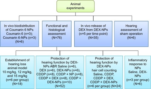
Figure 2 Characterization of DEX-NPs.
Notes: (A) Fabrication of DEX-NPs. Schematic representation of the nanoparticle structure: DEX (yellow balls) was encapsulated in the nanoparticles (light blue sphere) modified with PEG (gray ball shell). (B) Representative transmission electron microscopy image. (C) Two-dimensional nanoparticle image of atomic force microscopy. (D) Size distribution determined by dynamic light scattering. (E) In vitro release of DEX from DEX-NPs in PBS (pH 7.4), rat plasma, and AP (pH 7.4) (mean ± SD; n=3).
Abbreviations: AP, artificial perilymph; PBS, phosphate buffered saline; PEG, polyethylene glycol; d, days; DEX-NPs, dexamethasone-loaded polyethylene glycol-coated polylactic acid stealth nanoparticles; DEX, dexamethasone; SD, standard deviation; n, number.
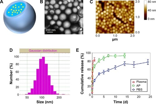
Figure 3 Coumarin 6-labeled NPs distribution in the cochlea via RWM administration at 1 hour after drug exposure.
Notes: (A) Weak fluorescence was detected in the cochlear modiolus after free Coumarin 6 RWM administration. Scale bar, 500 μm. Nuclei were stained with DAPI (blue). (B) Strong green fluorescence regions were visible in the stria vascularis and organs of Corti in all the cochlear turns, as well as in the spiral ganglion cells after Coumarin 6 NPs RWM administration.
Abbreviations: NP, nanoparticle; RWM, round window membrane; DAPI, 4′6-diamidino-2-phenylindole.
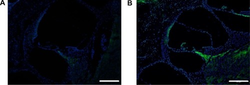
Figure 4 In vivo DEX concentrations in cochlear perilymph via RWM administration of free DEX or DEX-NPs.
Note: Data are presented as the mean ± SEM (n=5).
Abbreviations: DEX, dexamethasone; DEX-NPs, dexamethasone-loaded polyethylene glycol-coated polylactic acid stealth nanoparticles; h, hours; RWM, round window membrane; SEM, standard error of the mean; n, number.
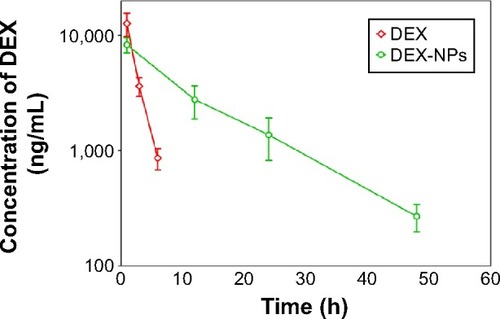
Figure 5 Establishment of cisplatin-induced hearing loss animal models.
Notes: Mean (A) ABR threshold and (B) shift per frequency for guinea pigs 3 days after treatment with 10 mg/kg and 12 mg/kg of intraperitoneal cisplatin (n=6). *P<0.05, **P<0.01, and ***P<0.001 as compared with baseline. ###P<0.001 as compared with the 10 mg/kg group.
Abbreviations: ABR, auditory brainstem response; SPL, sound pressure level; n, number.
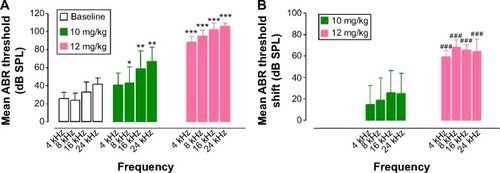
Figure 6 Average ABR threshold (mean ± SD; n=6) for each frequency prior to (0 d) and after (1 d and 3 d) the sham operation of the left ear.
Abbreviations: ABR, auditory brainstem response; SPL, sound pressure level; d, days; SD, standard deviation; n, number.
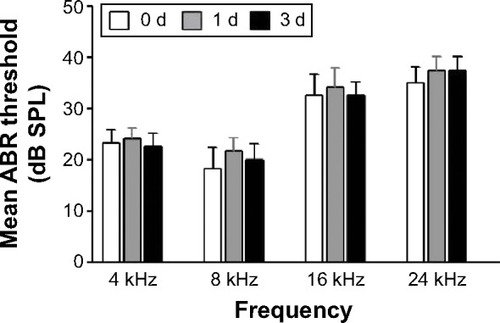
Figure 7 Mean ABR threshold and shift of guinea pigs treated with saline, DEX, DEX-NPs, CDDP, CDDP + NPs, CDDP + DEX, and CDDP + DEX-NPs by RWM administration.
Notes: Mean (A and C) ABR threshold and (B and D) shift of guinea pigs treated with saline (n=6), DEX (n=6), DEX-NPs (n=6), CDDP (n=8), CDDP + NPs (n=8), CDDP + DEX (n=8), and CDDP + DEX-NPs (n=10) by RWM administration. ABR shift thresholds are given as the mean ± SD. *P<0.05 and **P<0.01 as compared with CDDP; #P<0.05 and ##P<0.01 as compared with CDDP + DEX.
Abbreviations: ABR, auditory brainstem response; SPL, sound pressure level; DEX, dexamethasone; DEX-NPs, dexamethasone-loaded polyethylene glycol-coated polylactic acid stealth nanoparticles; CDDP, cis-diamminedichloroplatinumII; NP, nanoparticle; RWM, round window membrane; SD, standard deviation; n, number.
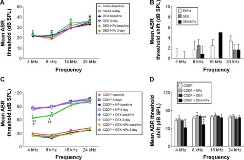
Figure 8 DEX-NPs alleviate cisplatin-induced damage on cochlear hair cells.
Notes: (A) Rhodamine phalloidin staining of the 60% portion from the apex of Corti. Severe damage to the OHCs was observed in the CDDP- and CDDP + DEX-treated animals, which were compared to CDDP + DEX-NPs. Scale bar, 40 μm. (B) Numbers and the percent survival of OHCs in 1 mm length about 60% from the apex of each cochlea (n=6).
***P<0.001 versus the CDDP group. #P<0.05, ###P<0.001 versus the CDDP + DEX group. Quantitative analyses showed that the DEX-NP treatment exhibited significant differences in the number and percentage of OHCs.
Abbreviations: OHCs, outer hair cells; IHCs, inner hair cells; CDDP, cis-diamminedichloroplatinumII; DEX, dexamethasone; DEX-NPs, dexamethasone-loaded polyethylene glycol-coated polylactic acid stealth nanoparticles; n, number.
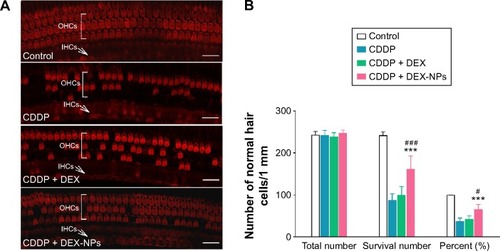
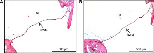
Figure 9 Hematoxylin and eosin-stained histological sections of a decalcified guinea pig middle-ear sample.
Notes: (A) RWM of the control ear with saline application. (B) RWM 1 day after the DEX-NPs administration. Blood is also visible in the scala tympani (n=3).
Abbreviations: RWM, round window membrane; ST, scala tympani; DEX-NPs, dexamethasone-loaded polyethylene glycol-coated polylactic acid stealth nanoparticles; n, number.