Figures & data
Figure 1 Schematic representation of active and passive targeting of DOX-loaded P407/FA-P407-TPGS micelles in cancer cells.
Notes: 1: DOX was loaded into micelles by solvent evaporation and thin-film hydration methods. 2: DOX-loaded micelles enter the cells by either passive transport or active transport mediated by FA ligand. Both active and passive transports across the cells are illustrated in enlarged version of cells. 3: Once inside the cells, DOX is released and acts on the target site to cause cell death. TPGS and P407 reduce drug efflux and enhance DOX–DNA binding. In both cases, that is, 3a and 3b, the ultimate goal is the cell death in which the targeted micelles result in enhanced cell death as compared to nontargeted DOX micelles.
Abbreviations: DOX, doxorubicin; P407, poloxamer 407; FA, folic acid; TPGS, D-α-tocopheryl polyethylene glycol succinate.
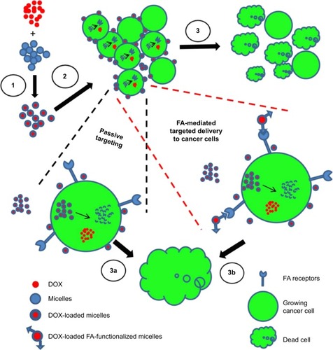
Figure 2 Synthesis of FA-P407 conjugate.
Notes: FA and P407 were conjugated by CDI-mediated coupling, purified by dialysis, and recovered by freeze drying.
Abbreviations: FA, folic acid; P407, poloxamer 407; CDI, 1,1′-carbonyldiimidazole; DMSO, dimethyl sulfoxide.
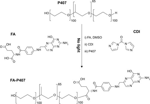
Figure 3 1H NMR spectra of FA, P407, and FA-P407.
Notes: Peaks a–c represent aromatic protons in FA, whereas peak d represents methyl protons of PPO confirming the conjugation of FA to P407.
Abbreviations: FA, folic acid; P407, poloxamer 407; PPO, polypropylene oxide.
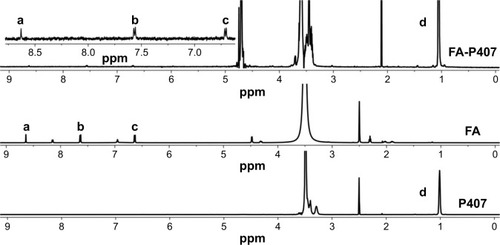
Figure 4 UV–vis spectra of FA, P407, and FA-P407.
Notes: FA and FA-P407 show characteristic absorption peaks at around 280 nm and 360 nm, whereas P407 has no peaks in this region.
Abbreviations: FA, folic acid; P407, poloxamer 407.
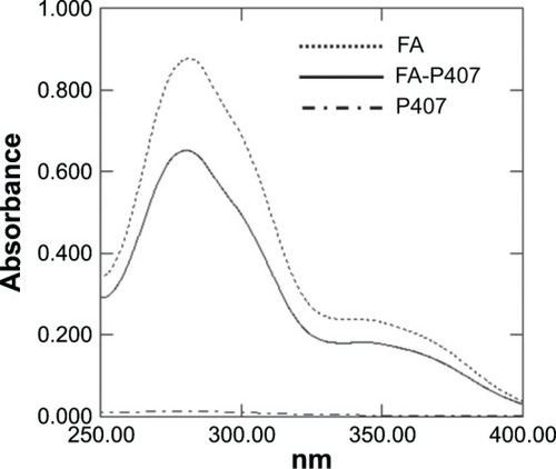
Table 1 Characterization of P407/FA-P407-TPGS micelles
Figure 5 Morphology of micelles.
Notes: (A) FESEM image of FA-P407-TPGS micelles. (B) TEM image of FA-P407-TPGS micelles.
Abbreviations: FESEM, field emission scanning electron microscopy; FA, folic acid; P407, poloxamer 407; TPGS, D-α-tocopheryl polyethylene glycol succinate; TEM, transmission electron microscopy.
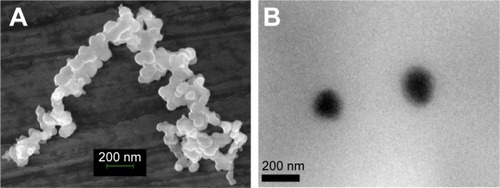
Table 2 Kinetic parameters of DOX release from micelles
Figure 6 pH-dependent DOX release from P407/FA-P407-TPGS micelles.
Notes: Red symbols: free DOX; symbols with lines: DOX release at pH 5; symbols without lines: DOX release at pH 7.
Abbreviations: DOX, doxorubicin; P407, poloxamer 407; FA, folic acid; TPGS, D-α-tocopheryl polyethylene glycol succinate.
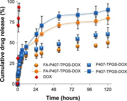
Figure 7 Nile red uptake.
Notes: SKOV3 cells were seeded on glass coverslips in 35 mm culture dishes. After 24 hours, cells were washed and treated with medium containing 0.5 μM nile red or equivalent concentration of nile red-loaded micelles. Nile red images were pseudo-colored green and merged with Hoechst 33342 images. Scale bars =50 μm.
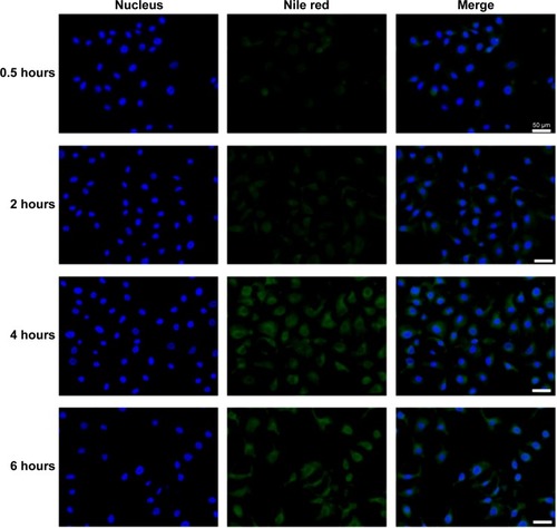
Figure 8 Drug efflux and DOX–DNA binding.
Notes: (A) R123 intracellular levels in both DOX-sensitive SKOV3 cells and SKOV3-DOX cells are shown after 2-hour treatment with TPGS, P407 alone, or P407-TPGS or FA-P407-TPGS micelles. (B) DOX binding with DNA in SKOV3 and SKOV3-DOX cells is shown as determined by fluorescence measurements.
Abbreviations: DOX, doxorubicin; R123, rhodamine 123; SKOV3-DOX, DOX-resistant SKOV3; TPGS, D-α-tocopheryl polyethylene glycol succinate; P407, poloxamer 407; FA, folic acid.
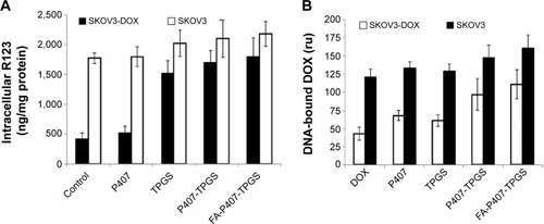
Figure 9 DOX uptake.
Notes: SKOV3 cells cultured on glass coverslips were treated with DOX for 4 hours. Images were acquired and merged with Hoechst 33342 images. Asterisk in the last row refers to cells cultured in the FA-free medium. Scale bars =50 μm.
Abbreviations: DOX, doxorubicin; FA, folic acid; P407, poloxamer 407; TPGS, D-α-tocopheryl polyethylene glycol succinate.
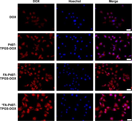
Table 3 IC50 values of DOX in free or micelle form
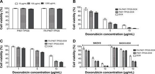
Figure 10 Cell viability of blank micelles and cytotoxicity of DOX and DOX-loaded micelles.
Notes: (A) Cell viability of WRL-68 cells after 24-hour treatment with P407-TPGS or FA-P407-TPGS micelles was determined by alamarBlue® cell viability assay. Cytotoxicity assessment of free DOX and DOX-loaded P407-TPGS or FA-P407-TPGS micelles by alamarBlue® assay is shown for (B) DOX-sensitive SKOV3 cells and (C) SKOV3-DOX cells. (D) Cytotoxicity of DOX or DOX-loaded micelles in SKOV3 and SKOV3-DOX cells in medium supplemented with 2 mM FA.
Abbreviations: DOX, doxorubicin; P407, poloxamer 407; TPGS, D-α-tocopheryl polyethylene glycol succinate; FA, folic acid; SKOV3-DOX, DOX-resistant SKOV3.