Figures & data
Figure 1 The characterization of gold nanoclusters (AuNCs) and RDP-AuNCs.
Notes: (A) Appearance and electrophoretic analysis of AuNCs and FAM-RDP-AuNCs. Aqueous solution and powder under visible and ultraviolet light (365 nm). (a) AuNCs; (b) FAM-RDP-AuNCs. The left figure is aqueous solution, the middle is powder under visible light, and the right figure is powder under ultraviolet light. (B) Electrophoretic mobility of AuNCs (Lane 1) and FAM-RDP-AuNCs (Lane 2) in agarose gel. (C) Size distribution of AuNCs and RDP-AuNCs determined by dynamic light scattering. (D) Fourier-transform infrared spectra of AuNCs and RDP-AuNCs. (E) Fluorescence of AuNCs and RDP-AuNCs (λex=510 nm). (F) Absorption spectra of AuNCs and RDP-AuNCs. Red curve: AuNCs; green curve: RDP-AuNCs.
Abbreviations: AuNCs, gold nanoclusters; FAM, carboxyfluorescein; RDP, rabies virus glycoprotein derived peptide.
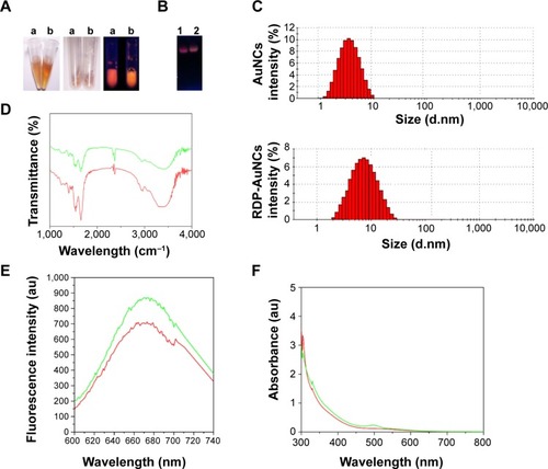
Figure 2 Cellular uptake analysis.
Notes: (A) Fluorescence images of SH-SY5Y cells treated with gold nanoclusters (AuNCs) or RDP-AuNCs. Nuclei (blue) were stained with 4′,6-diamidino-2-phenylindole (DAPI). (B) Cellular uptake specificity of AuNCs (red) conjugated with FAM-RDP (green) into neural cells. (C) Time course of intracellular accumulation of FAM-labeled RDP-AuNCs observed under fluorescence microscopy.
Abbreviation: AuNCs, gold nanoclusters; FAM, carboxyfluorescein; RDP, rabies virus glycoprotein derived peptide.
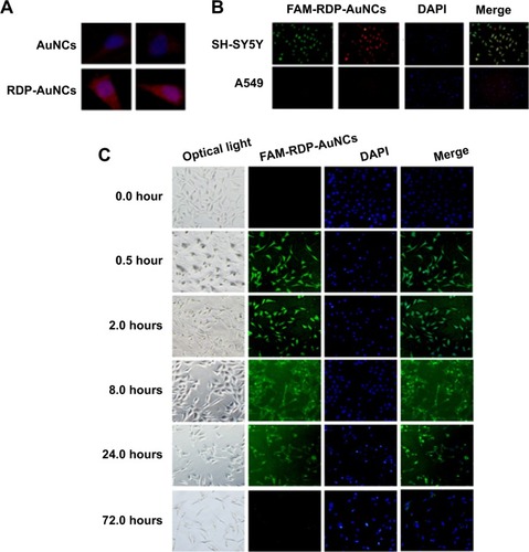
Figure 3 Cellular uptake of FAM-RDP-AuNCs in gliomas cell lines (U87) after 8 hours’ incubation. Nuclei were stained with 4′,6-diamidino-2-phenylindole (DAPI).
Abbreviations: AuNCs, gold nanoclusters; FAM, carboxyfluorescein; RDP, rabies virus glycoprotein derived peptide.
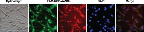
Figure 4 In vitro cytotoxicity assay of RDP-AuNCs in (A) SH-SY5Y cells and (B) U87 cells by 3-(4,5-dimethylthiazol-2-yl)-2,5-diphenyltetrazolium bromide (MTT) assay.
Note: Data are expressed as mean ± standard error of the mean (n=4).
Abbreviations: AuNCs, gold nanoclusters; RDP, rabies virus glycoprotein derived peptide.
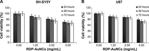
Figure 5 Internalization mechanism of RDP-AuNCs.
Notes: (A) Pretreatment with ATP-depletion buffer; (B) treatment with K+-depletion buffer. In (A,B) the arrows point to the attachment of RDP-AuNCs on the cell membrane; (C) normal; (D) RDP-AuNCs entrapped in the endosome under transmission electron microscopy (TEM). The arrows point to the RDP-AuNCs trapped in the endosome. (E) no endosome was observed in the cells after 2 hours. The arrows point to the RDP-AuNCs dispersed in the cytoplasm.
Abbreviations: AuNCs, gold nanoclusters; ATP, adenosine triphosphate; N, nucleus under TEM; RDP, rabies virus glycoprotein derived peptide.
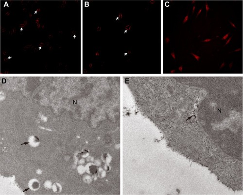
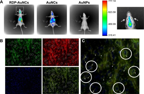
Figure 6 In vivo imaging of RDP-AuNCs.
Notes: (A) Noninvasive imaging 1 hour after intravenous (IV) injection into the tail vein of RDP-AuNCs, gold nanoclusters (AuNCs), and gold nanoparticles (AuNPs), respectively. (B) The mouse brain was dissected to take slices at 2 hours after IV injection with FAM-RDP-AuNCs. Green spots represent the fluorescence from FAM of FAM-RDP-AuNCs; red spots represent fluorescence from nanoclusters of FAM-RDP-AuNCs; 4′,6-diamidino-2-phenylindole (DAPI)-stained nuclei appear blue. Merge of images; (C) magnification of circles of merge images. The arrows point to FAM-RDP-AuNC fluorescence.
Abbreviations: AuNCs, gold nanoclusters; FAM, carboxyfluorescein; RDP, rabies virus glycoprotein derived peptide.