Figures & data
Figure 1 Schematic representation of the design of this study.
Note: Synthesis of M-MSN_siRNA@PEI-PEG-KALA (top) and systemic administration of VEGF siRNA via this nanocarrier into orthotopic ovarian tumor-bearing mice led to effective silencing of VEGF gene expression in cancer cells and inhibited angiogenesis, ultimately leading to suppression of cancer growth.
Abbreviations: M-MSN, magnetic mesoporous silica nanoparticle; siRNA, small interfering RNA; PDI, polydispersity index; PEI, polyethylenimine; PEG, polyethylene glycol; KALA, a type of fusogenic peptide; VEGF, vascular endothelial growth factor.
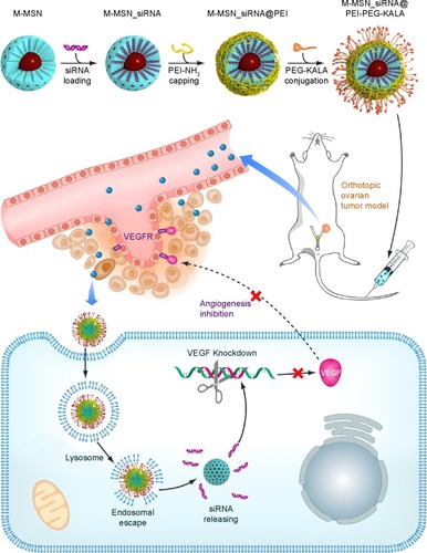
Figure 2 (A) Transmission electron microscopic image of M-MSN_siRNA@PEI-PEG-KALA. (B) Dynamic light scattering measurements of size distribution for M-MSN_siRNA@PEI-PEG-KALA (dispersed in saline). (C) Zeta potential of M-MSN_siRNA@PEI-PEG-KALA. (D) T2 relaxation rate (1/T2) as a function of iron concentration for M-MSN_siRNA@PEI-PEG-KALA.
Notes: Scale bar, 20 nm. (B) The polydispersity index (PDI) of this nanocarrier was 0.095.
Abbreviations: M-MSN, magnetic mesoporous silica nanoparticle; siRNA, small interfering RNA; PEI, polyethylenimine; PEG, polyethylene glycol; KALA, a type of fusogenic peptide.
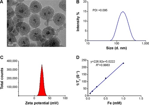
Figure 3 (A) An MTT assay was performed to evaluate the viability of the SKOV3 cells on exposure to M-MSN_NC siRNA@PEI-PEG-KALA containing various concentrations of M-MSNs, ranging from 50 to 400 μg/mL. (B) Downregulated vascular endothelial growth factor (VEGF) expression in SKOV3 cells by the M-MSN_VEGF siRNA@PEI-PEG-KALA, M-MSN_VEGF siRNA@PEI and M-MSN_VEGF siRNA@PEI-PEG delivery systems.
Notes: (A) 0 denotes absence of treatment. (B) The dose of siRNA(NCor VEGF) was 150 nM within 80 μg/mL of M-MSNs. An identical quantity of siRNA was also transfected with Lipofectamine™ 2000 (***P<0.0001, n=3).
Abbreviations: MTT, 3-(4,5-dimethylthiazol-2-yl)-2,5-diphenyltetrazolium bromide; M-MSN, magnetic mesoporous silica nanoparticle; PEI, polyethylenimine; PEG, polyethylene glycol; KALA, a type of fusogenic peptide; VEGF siRNA, vascular endothelial growth factor small interfering RNA; NC siRNA, negative control small interfering RNA.
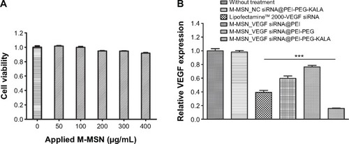
Figure 4 (A) Orthotopic ovarian mouse tumor (red circle is the tumor position). (B) In vivo T2-weighted magnetic resonance images of the xenograft tumor model before (left) and 24 hours after (right) injection of M-MSN_NC siRNA@PEI-PEG-KALA.
Notes: Gray-scale images (top, I and II) and pseudocolor images (bottom, I’ and II’) are shown.
Abbreviations: M-MSN, magnetic mesoporous silica nanoparticles; PEI, polyethylenimine; PEG, polyethylene glycol; KALA, a type of fusogenic peptide; NC siRNA, negative control small interfering RNA.
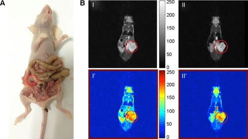
Figure 5 (A) Changes in body weight in the different treatment groups. (B) Tumors collected from the four treatment groups at the end of the experiment. Tumor weights (C) and tumor volumes (D) were measured after euthanizing the mice.
Notes: Data represent the mean ± standard deviation. (*P<0.01; **P<0.001; ***P<0.0001; n=5). a: M-MSN_VEGF siRNA@PEI-PEG-KALA; b: M-MSN_NC siRNA@PEI-PEG-KALA; c: M-MSN@PEI-PEG-KALA; d: saline.
Abbreviations: M-MSN, magnetic mesoporous silica nanoparticles; PEI, polyethylenimine; PEG, polyethylene glycol; KALA, a type of fusogenic peptide; NC siRNA, negative control small interfering RNA; VEGF siRNA, vascular endothelial growth factor small interfering RNA.
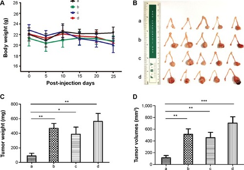
Figure 6 (A) Immunohistological analysis for VEGF expression in intratumoral sections (top) and microvessel density represented by CD31-positive endothelial cells (bottom) in the four treatment groups. (B) Intratumoral VEGF expression was measured by enzyme-linked immunosorbent assay. Statistical data reflect VEGF (C) and CD31 (D) expression in tumoral sections, respectively.
Notes: Data represent the mean ± standard deviation. (**P<0.001; ***P<0.0001; n=5). Magnification, 20×. a: M-MSN_VEGF siRNA@PEI-PEG-KALA; b: M-MSN_NC siRNA@PEI-PEG-KALA; c: M-MSN@PEI-PEG-KALA; d: saline.
Abbreviations: IOD, integral optical density; VEGF, vascular endothelial growth factor; M-MSN, magnetic mesoporous silica nanoparticle; MVD, microvessel density; PEI, polyethylenimine; PEG, polyethylene glycol; KALA, a type of fusogenic peptide.
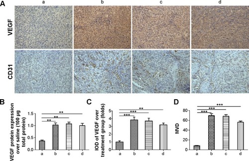
Figure S1 (A) TGA curves for various types of M-MSNs loaded with siRNA. (B) The standard curve for FITC-labeled KALA. Fluorescence intensity values at 520 nm versus the amount of FITC-labeled KALA.
Note: A linear fit was used for these data.
Abbreviations: TGA, thermogravimetric analysis; M-MSNs, magnetic mesoporous silica nanoparticles; FITC, fluorescein isothiocyanate; NC, negative control; PEI, polyethylenimine; PEG, polyethylene glycol; siRNA, small interfering RNA; KALA, a type of fusogenic peptide; TG, thermogravimetry.
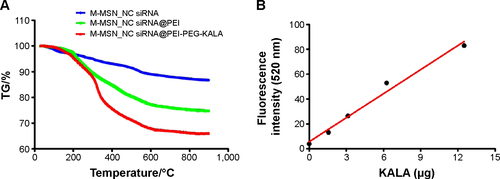
Figure S2 An MTT assay was performed to evaluate the viability of the SKOV3 cells by M-MSN_NC siRNA@PEI with various concentrations of applied M-MSNs ranging from 50 to 400 μg/mL.
Notes: 0 denotes absence of treatment. *P<0.01; **P<0.001; ***P<0.0001, n=3.
Abbreviations: MTT, 3-(4,5-dimethylthiazol-2-yl)-2,5-diphenyltetrazolium bromide; M-MSN, magnetic mesoporous silica nanoparticle; PEI, polyethylenimine; NC siRNA, negative control small interfering RNA.
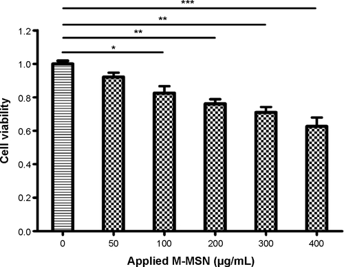
Figure S3 Fluorescent confocal laser scanning microscopic images of SKOV3 cells after incubation with M-MSN_NC siRNA@PEI-PEG-KALA nanocarriers for 24 hours (A, B) or 36 hours (C).
Notes: (AI) Blue fluorescent image of cell nucleus stained by DAPI, (AII) green fluorescent image of siRNA labeled by FAM, (AIII) differential interference contrast image, and (AIV) overlaying image of I–III. (BI) Green fluorescent images of FAM-labeled siRNA, (BII) red fluorescent images of endolysosomes stained by LysoTracker® Red DND, (BIII) merged image of I and II, and (BIV) differential interference contrast image. (CI) Green fluorescent images of FAM-labeled siRNA, (CII) red fluorescent images of M-MSNs labeled by cyanine 5.5, (CIII) merged image of I and II, and (CIV) differential interference contrast image. Magnification 20×.
Abbreviations: DAPI, 4′,6-diamidino-2-phenylindole; FAM, fluorescein amidite; M-MSN, magnetic mesoporous silica nanoparticle; PEI, polyethylenimine; PEG, polyethylene glycol; KALA, a type of fusogenic peptide; siRNA, small interfering RNA.
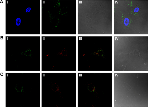
Figure S4 Flow cytometry results for internalization ability among the various M-MSN-based siRNA delivery systems 4 hours after incubation with SKOV3 cells.
Notes: Red line indicates without treatment with any type of particle; green line indicates M-MSN_FAM-siRNA@PEI-PEG; black line indicates M-MSN_FAM-siRNA@PEI; and blue line indicates M-MSN_FAM-siRNA@PEI-PEG-KALA.
Abbreviations: FAM, fluorescein amidite; FITC, fluorescein isothiocyanate; M-MSN, magnetic mesoporous silica nanoparticle; PEI, polyethylenimine; PEG, polyethylene glycol; KALA, a type of fusogenic peptide; siRNA, small interfering RNA.
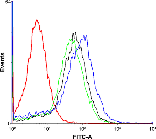
Figure S5 Iron from M-MSNs was revealed by Prussian blue staining.
Notes: (A) Tumor slide after injection of M-MSN_NC siRNA@PEI-PEG-KALA (red arrow points to the iron accumulated in tumor region). (B) Liver tissue from animals that were not injected with nanocarriers, but the equivalent saline. Magnification was 20×.
Abbreviations: M-MSN, magnetic mesoporous silica nanoparticles; PEI, polyethylenimine; PEG, polyethylene glycol; KALA, a type of fusogenic peptide; NC siRNA, negative control small interfering RNA.
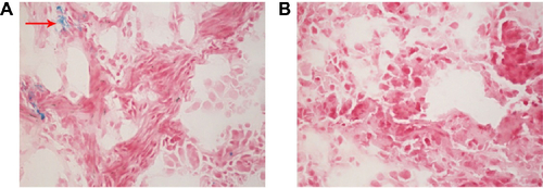
Figure S6 Safety and biocompatibility of M-MSN_VEGF siRNA@PEI-PEG-KALA.
Notes: Representative markers of nephrotoxicity and hepatotoxicity were evaluated, including ALT (A), AST (B), urea (C), creatinine (D), and serum uric acid (E). a: M-MSN_VEGF siRNA@PEI-PEG-KALA; b: M-MSN_NC siRNA@PEI-PEG-KALA; c: M-MSN@PEI-PEG-KALA; d: saline.
Abbreviations: ALT, alanine aminotransferase; AST, aspartate aminotransferase; M-MSN, magnetic mesoporous silica nanoparticle; PEI, polyethylenimine; PEG, polyethylene glycol; KALA, a type of fusogenic peptide; VEGF siRNA, vascular endothelial growth factor small interfering RNA.
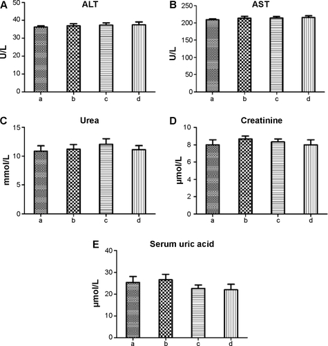
Figure S7 Hematoxylin and eosin staining of the major organs, including the heart, liver, spleen, lung, and kidney.
Notes: Magnification 20×. A: M-MSN_VEGF siRNA@PEI-PEG-KALA; B: M-MSN_NC siRNA@PEI-PEG-KALA; C: M-MSN@PEI-PEG-KALA; D: saline.
Abbreviations: M-MSN, magnetic mesoporous silica nanoparticle; NC, negative control; PEI, polyethylenimine; PEG, polyethylene glycol; KALA, a type of fusogenic peptide; VEGF siRNA, vascular endothelial growth factor small interfering RNA.
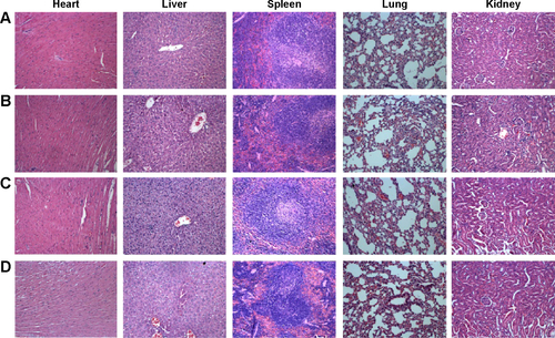
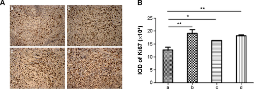
Figure S8 (A) Immunohistological analysis for Ki-67 expression in tumor sections from the four experimental groups. (B) Statistical analysis from immunohistological results.
Notes: Data are shown as the mean ± standard deviation. a: M-MSN_VEGF siRNA@PEI-PEG-KALA; b: M-MSN_NC siRNA@PEI-PEG-KALA; c: M-MSN@PEI-PEG-KALA; d: saline. (*P<0.01; **P<0.001; n=5). Magnification 20×.
Abbreviation: IOD, integral optical density.