Figures & data
Figure 1 Characterization of HAP sol.
Notes: (A) Photograph of HAP precursor sol, (B) TEM micrograph of HAP nanoparticles.
Abbreviations: HAP, hydroxyapatite; TEM, transmission electron microscopy.
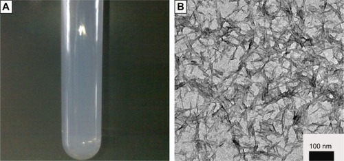
Figure 2 XRD pattern of HAP particles in sol.
Abbreviations: HAP, hydroxyapatite; XRD, X-ray diffraction.
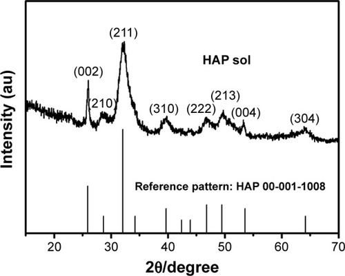
Figure 3 The morphology of electrospun fibers with different HAP contents.
Notes: (A) 0%, (B) 10%, (C) 20%, (D) 30%, (E) 40%, and collagen/30% HAP control fibers (F), respectively.
Abbreviation: HAP, hydroxyapatite.
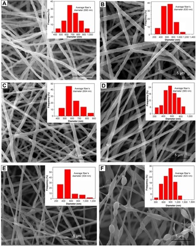
Figure 4 TEM images of the fibers and spinning solutions.
Notes: (A) Collagen fibers before desalination, (B) collagen/HAP before desalination, (C) collagen fibers after desalination, (D) collagen/HAP after desalination, (E) collagen/30% HAP spinning solution, (F) collagen/30% HAP fibers after desalination, (G) collagen/HAP spinning solution using HAP powders, (H) collagen/30% HAP control fibers.
Abbreviations: HAP, hydroxyapatite; TEM, transmission electron microscopy.
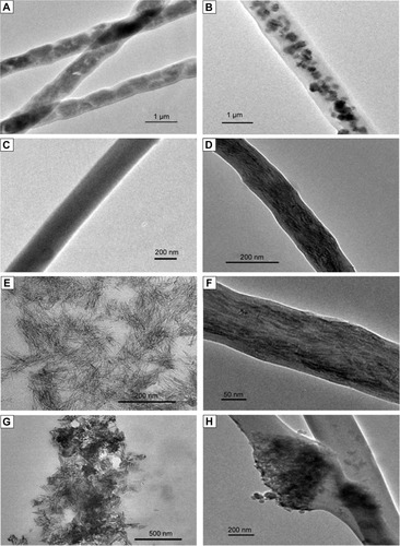
Figure 5 Schematic illustration of the proposed mechanism for the HAP particles oriented along the long axes of the collagen fiber.
Abbreviation: HAP, hydroxyapatite.
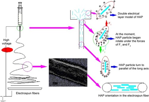
Figure 6 FTIR spectra.
Notes: (A) HAP, (B) collagen fibers, (C) collagen/HAP composite fibers.
Abbreviations: HAP, hydroxyapatite; FTIR, Fourier-transform infrared spectroscopy; au, arbitrary units.
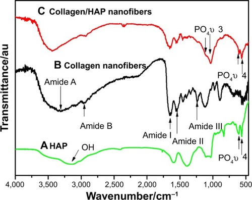
Figure 7 XRD patterns of collagen fibers and collagen/HAP fibers.
Notes: (A) Collagen fibers before desalination, (B) collagen fibers after desalination, (C) collagen/30% HAP composite fibers.
Abbreviations: HAP, hydroxyapatite; XRD, X-ray diffraction.
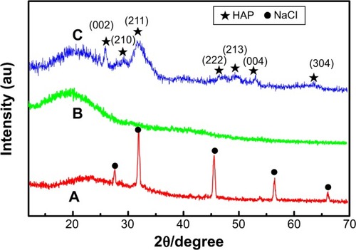
Figure 8 SEM photographs of the electrospun fibers after cross-linking with EDC/NHS.
Notes: (A) Collagen fibers, (B) collagen/10% HAP fibers, (C) collagen/30% HAP fibers.
Abbreviations: HAP, hydroxyapatite; SEM, scanning electron microscope; EDC/NHS, 1-ethyl-3-(3-dimethyl-aminopropyl)-1-carbodiimide hydrochloride/N-hydroxysuccinimide.
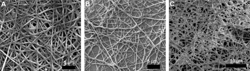
Table 1 Mechanical properties of collagen and collagen/HAP composite fibers with different content of HAP
Figure 9 Viability of U2-OS cells in leaching liquors of collagen and collagen/HAP composite nanofibers after culture for 1, 3, 5, and 7 days.
Abbreviations: HAP, hydroxyapatite; PTFE, polytetrafluoroethylene; d, day(s).
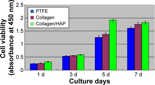
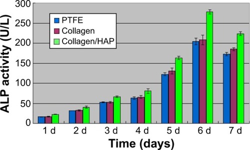
Figure 10 ALP activity of U2-OS cells after being cultured for 1, 2, 3, 4, 5, 6, and 7 days in medium containing material extraction.
Abbreviations: HAP, hydroxyapatite; PTFE, polytetrafluoroethylene; d, day(s).