Figures & data
Table 1 Physicochemical properties of the calcium carbonates examined
Figure 1 XRD patterns and SEM images of B-Cal (a) and N-Cal (b) as produced (A) and after being incubated in simulated gastric (B) and intestinal fluids (C) and plasma (D).
Notes: (hkl) Miller indexing in XRD patterns (A) are typical patterns of the calcite phase (JCPDS No 47-1743); Asterisks in XRD patterns stand for the evolution of dicalcium phosphate dihydrate (JCPDS No 72-0714); SEM images were obtained after incubation for 1 hour in simulated gastric fluid and 4 hours in simulated intestinal fluid or plasma condition.
Abbreviations: arb, arbitrary; B-Cal, bulk calcium carbonates; hkl, Miller indexing; JCPDS, Joint Committee on Powder Diffraction Standards; N-Cal, nano calcium carbonates; SEM, scanning electron microscopy; XRD, X-ray diffraction; h, hours.
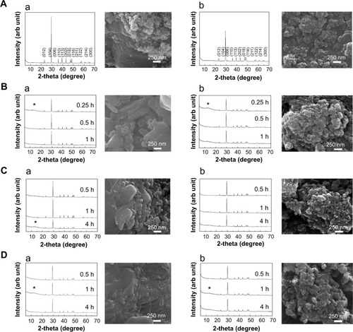
Table 2 In vitro and ex vivo dissolution and protein fluorescence quenching ratio of calcium carbonate materials
Table 3 List of the most abundant plasma proteins bound on B-Cal and N-Cal as determined by LC–MS
Figure 2 Plasma protein binding profiles of calcium carbonates separated by one-dimensional (A), and two-dimensional (B), gel electrophoresis.
Abbreviations: B-Cal, bulk calcium carbonates; N-Cal, nano calcium carbonates.
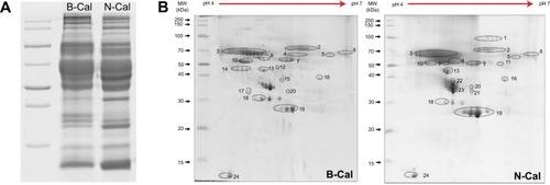
Table 4 Biokinetic parameters and absorption of calcium carbonates after oral administration to rats
Figure 3 Plasma concentration-time curves (A–D) and tissue distribution (E, F) of calcium carbonates after a single (A, B), or 14 days of daily oral treatment (C–F) to rats.
Notes: Biokinetic data are presented as increase in calcium levels after subtracting the basal plasma calcium levels observed in untreated controls; (A) Biokinetics after administering a single dose to male rats; (B) Biokinetics after administering a single dose to female rats; (C) Biokinetics after 14-days of daily oral treatment in male rats; (D) Biokinetics after 14-days of daily oral treatment in female rats; (E) Tissue distribution in male rats; (F) Tissue distribution in female rats. (a) and (b) is used when there is no significant difference between two groups (control and N-Cal/N-Cal and B-Cal), but significant difference is found when three groups (control, N-Cal, B-Cal) are compared.
Abbreviations: B-Cal, bulk calcium carbonates; N-Cal, nano calcium carbonates.
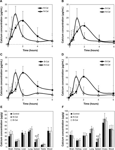
Figure S1 XRD patterns (A) and FT-IR spectra (B) of (a) B-Cal and (b) N-Cal.
Abbreviations: arb, arbitrary; B-Cal, bulk calcium carbonates; FT-IR, Fourier transform infrared; N-Cal, nano calcium carbonates; XRD, X-ray diffraction.
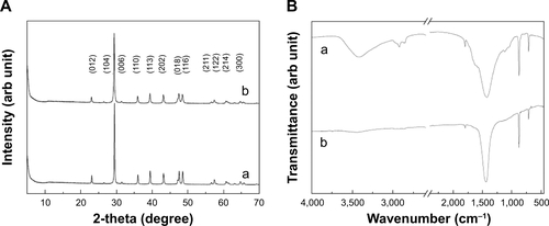
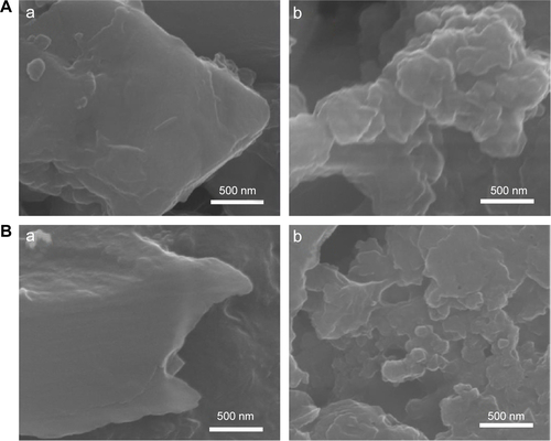
Figure S2 SEM images for the produced B-Cal (A, C) and N-Cal (B, D).
Abbreviations: B-Cal, bulk calcium carbonates; N-Cal, nano calcium carbonates; SEM, scanning electron microscopy.
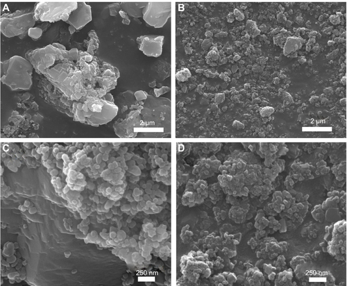
Figure S3 Hydrodynamic size distributions of B-Cal (A), and N-Cal (B) in aqueous suspension as determined by dynamic light scattering.
Abbreviations: B-Cal, bulk calcium carbonates; N-Cal, nano calcium carbonates; PDI, polydispersity index
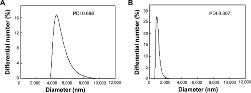
Figure S4 Zeta potential distributions of B-Cal (A), and N-Cal (B).
Abbreviations: arb, arbitrary; B-Cal, bulk calcium carbonates; N-Cal, nano calcium carbonates.
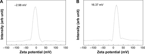
Figure S5 XRD patterns of B-Cal (a), and N-Cal (b), incubated in simulated body fluids: gastric fluid (A), intestinal fluid (B), and plasma (C) after 24 or 48 hours.
Note: Asterisks indicate the evolution of dicalcium phosphate dihydrate (JCPDS No 72-0714).
Abbreviations: arb, arbitrary; B-Cal, bulk calcium carbonates; JCPDS, Joint Committee on Powder Diffraction Standards; N-Cal, nano calcium carbonates; XRD, X-ray diffraction; h, hours.
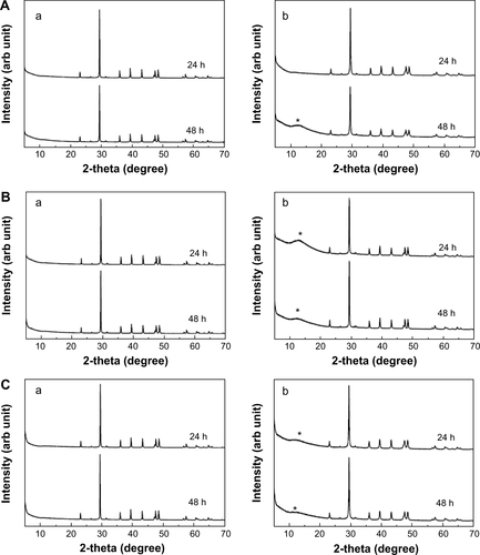
Figure S6 XANES at calcium K-edge for B-Cal (A), and N-Cal (B), treated with simulated body fluid, untreated (a) and gastric fluid (b), intestinal fluid (c) and plasma (d) for 48 hours.
Note: The inset table shows the edge position obtained by secondary differentiation of peak.
Abbreviations: arb, arbitrary; B-Cal, bulk calcium carbonates; N-Cal, nano calcium carbonates; XANES, X-ray absorption near edge structure.
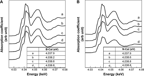
Figure S7 XRD patterns of B-Cal (a) and N-Cal (b) incubated ex vivo in fluids extracted from tissues: gastric fluid (A), intestinal fluid (B), and plasma (C).
Note: Asterisks indicate the evolution of dicalcium phosphate dihydrate (JCPDS No 72-0714).
Abbreviations: arb, arbitrary; B-Cal, bulk calcium carbonates; JCPDS, Joint Committee on Powder Diffraction Standards; N-Cal, nano calcium carbonates; XRD, X-ray diffraction; h, hours.
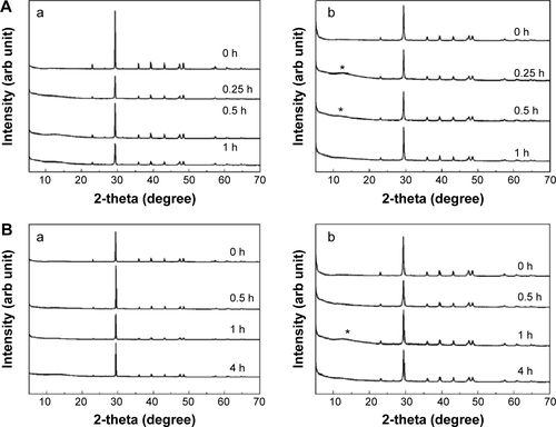
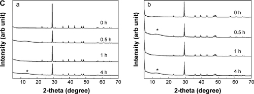
Figure S8 SEM images of B-Cal (a) and N-Cal (b) after incubation ex vivo in tissue extracted fluids: gastric fluid for 1 hour (A), intestinal fluid for 4 hours (B), and plasma for 4 hours (C).
Abbreviations: B-Cal, bulk calcium carbonates; N-Cal, nano calcium carbonates; SEM, scanning electron microscopy.
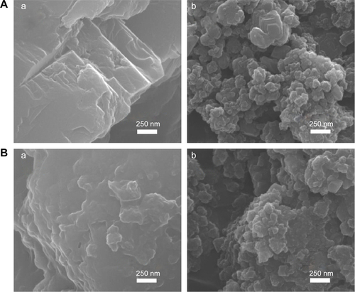
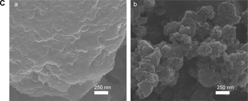
Figure S9 XRD patterns of B-Cal (a), and N-Cal (b), obtained from stomach (A), and intestine (B) following oral administration to rats.
Note: Asterisks and triangles indicate dicalcium phosphate dihydrate (JCPDS No 72-0714) and calcite, respectively.
Abbreviations: arb, arbitrary; B-Cal, bulk calcium carbonates; JCPDS, Joint Committee on Powder Diffraction Standards; N-Cal, nano calcium carbonates; XRD, X-ray diffraction; h, hours.
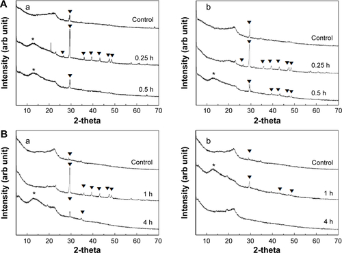
Figure S10 SEM images of B-Cal (a) and N-Cal (b) obtained from stomach at 1 hour postadministration (A), and intestine at 4 hours postadministration (B).
Abbreviations: B-Cal, bulk calcium carbonates; N-Cal, nano calcium carbonates; SEM, scanning electron microscopy.