Figures & data
Figure 1 Characterization of gold nanoparticles, VGC, and VGC-L.
Notes: (A) Particle size, zeta potential, encapsulation efficiency, and drug loading (n=3); (B) UV-visible spectra; (C) TEM images of gold nanoparticles (×100,000 magnification); (D) TEM images of VGC (×100,000 magnification); (E) TEM images of VGC-L (×100,000 magnification).
Abbreviations: PDI, polydispersity index; EE, Encapsulation efficiency; DL, drug loading; VGC-L, vincristine sulfate-gold nanoparticles conjugates-loaded liposomes; TEM, transmission electron microscopy; VGC, vincristine sulfate-gold nanoparticles conjugates; VCR, vincristine sulfate.
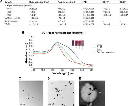
Figure 2 Characterization of VGC.
Notes: (A) UV/visible spectra of VGC at various pH; (B) UV/visible spectra of VGC at various NaCl concentrations; (C) VCR release profile from VGC in phosphate buffer (pH 7.4) and acetate buffer (pH 4.5).
Abbreviations: VGC, vincristine sulfate-gold nanoparticles conjugates; VCR, vincristine sulfate.
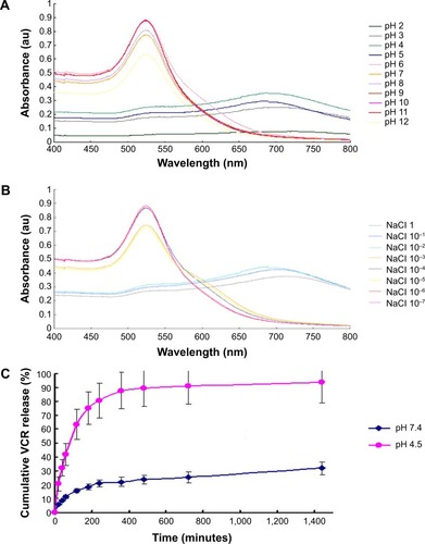
Figure 3 The drug release profile of VCR from VGC-L.
Notes: The drug release as measured in (A) in phosphate buffer (pH 7.4) and acetate buffer (pH 4.5); (B) in acetate buffer (pH 4.5) under 365-nm UV light.
Abbreviation: VCR, vincristine sulfate; VGC-L, vincristine sulfate-gold nanoparticles conjugates loaded liposomes.
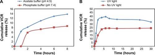
Figure 4 Cell uptake and cytotoxicity of VGC-L to Hela cells.
Notes: (A) TEM images of HeLa cells (×30,000 magnification). The red circles (1–6) represent the location of gold nanoparticles, the solid atrous dots in the circles are the gold nanoparticles, and the two arrows indicate the local area magnification; (B) The inhibition of HeLa cell viability following exposure to free VCR, VCR-L, and VGC-L with UV light for the indicated time periods.
Abbreviations: TEM, transmission electron microscopy; VCR, vincristine sulfate; VCR-L, vincristine sulfate-loaded liposomes; VGC-L, vincristine sulfate-gold nanoparticles conjugates loaded liposomes.
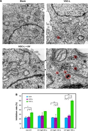
Table 1 Mean fluorescent intensity of HeLa cells exposed to liposomes loaded with calcein-conjugated gold nanoparticles (CGC-L) and CGC-L plus UV light, as determined by flow cytometry
Figure 5 Confocal laser scanning microscopy images of HeLa cells treated for the indicated times with liposomes loaded with CGC-L or CGC-L plus UV light exposure.
Notes: Column A, 0.5 h; B, 0.5 h + UV light; C, 1 h; D, 1 h + UV light; E, 2 h; F, 2 h + UV light. Row 1, Hoechst 33258 (blue); 2, Lyso-Tracker Red (red); 3, calcein (green); 4, merged.
Abbreviation: CGC-L, calcein-conjugated gold nanoparticles loaded liposomes.
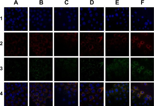
Figure 6 In vivo antitumor study in BALB/c mice implanted with HeLa cells.
Notes: (A) Body weight changes in control mice and those treated with UV laser only, VCR, VGC-L, and VGC-L plus UV laser (n=5). **P<0.01. (B) Relative tumor volume in control mice and those treated with UV laser only, VCR, VGC-L, and VGC-L plus UV laser (n=5). *P<0.05, **P<0.01. (C) Photographs of tumors after excision from (a) control; (b) only UV laser; (c) VCR; (d) VGC-L; and (e) VGC-L plus UV laser groups. (D) TUNEL staining of the isolated tumor tissue from (a) control; (b) blank plus UV laser; (c) VCR; (d) VGC-L; and (e) VGC-L plus UV laser groups. The apoptotic cells and cell nuclei were stained green and blue, respectively.
Abbreviation: VCR, vincristine sulfate; VGC-L, vincristine sulfate-gold nanoparticles conjugates loaded liposomes; TUNEL, Terminal-deoxynucleoitidyl Transferase Mediated Nick End Labeling.
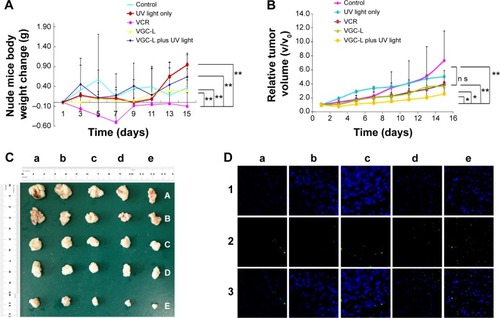
Figure 7 In vivo images of tumor-bearing nude mice treated with rhodamine B-conjugated gold nanoparticle-loaded liposomes (RGC-L) and RGC-L plus UV light.
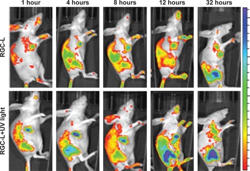
Figure S1 (A) FTIR spectra of (a) gold nanoparticles; (b) VCR; and (c) VGC. (B) DSC curves of (a) gold nanoparticles; (b) VCR; and (c) VGC. (C) DSC curves of (a) BL; (b) VGC; (c) VGC-L.
Abbreviations: FTIR, fourier transform infrared spectroscopy; VCR, vincristine sulfate; VGC, vincristine sulfate-gold nanoparticles conjugates; DSC, differential scanning calorimetry; BL, blank liposomes; VGC-L, vincristine sulfate-gold nanoparticles conjugates loaded liposomes.
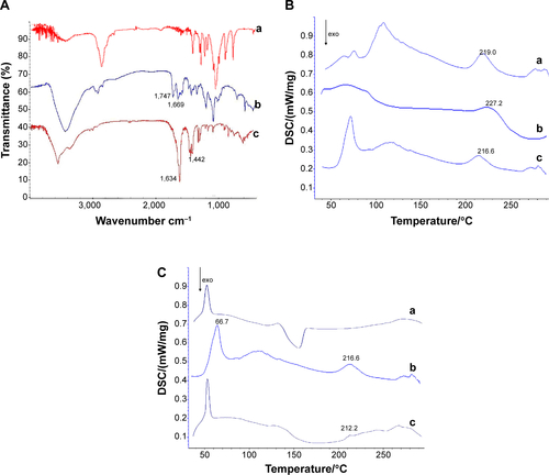
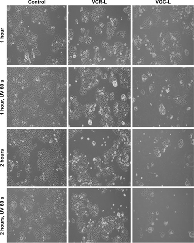
Figure S2 Microscope images of control HeLa cells and those incubated with VCR-L, VGC-L, and VGC-L plus UV light.
Abbreviations: VCR-L, vincristine sulfate-loaded liposomes; VGC-L, vincristine sulfate-gold nanoparticles conjugates loaded liposomes.