Figures & data
Figure 1 Preparation of DOX-LTSL-CREKA.
Notes: The schematic representation of synthesis of DSPE-PEG-CREKA (A). DOX-LTSL-CREKA, composed of DPPC, MSPC, DSPE-PEG, and DSPE-PEG-CREKA (86:10:2:2 molar ratio), was prepared by a thin-film hydration method (B). The DOX/lipid (w/w) ratio was 1:20. The schematic representation depicting the central concept of DOX-LTSL-CREKA (C). The encapsulated DOX was in the thermosensitive liposomes at 37°C. The encapsulated DOX immediately released from thermosensitive liposomes during hyperthermia treatment (43°C). Typical particle size and distribution of the DOX-LTSL-CREKA (D). The particle size of DOX-LTSL-CREKA during hyperthermia treatment (43°C) for 10 minutes (E).
Abbreviations: DOX, doxorubicin; LTSL, lysolipid-containing thermosensitive liposome; CREKA, Cys-Arg-Glu-Lys-Ala; DSPE-PEG, 1,2-distearoyl-sn-glycero-3-phos phatidylethanolamine-N-[methoxy(polyethylene glycol)-2000]; DPPC, 1,2-dipalmitoyl-sn-glycero-3-phosphatidylcholine; MSPC, mono-stearoyl-sn-glycero-3-phos-phatidylcholine; DSPE-PEG-MAL, 1,2-distearoyl-sn-glycero-3-phosphoethanolamine-N-[maleimide(polyethylene glycol)-2000]; PBS, phosphate-buffered saline.
![Figure 1 Preparation of DOX-LTSL-CREKA.Notes: The schematic representation of synthesis of DSPE-PEG-CREKA (A). DOX-LTSL-CREKA, composed of DPPC, MSPC, DSPE-PEG, and DSPE-PEG-CREKA (86:10:2:2 molar ratio), was prepared by a thin-film hydration method (B). The DOX/lipid (w/w) ratio was 1:20. The schematic representation depicting the central concept of DOX-LTSL-CREKA (C). The encapsulated DOX was in the thermosensitive liposomes at 37°C. The encapsulated DOX immediately released from thermosensitive liposomes during hyperthermia treatment (43°C). Typical particle size and distribution of the DOX-LTSL-CREKA (D). The particle size of DOX-LTSL-CREKA during hyperthermia treatment (43°C) for 10 minutes (E).Abbreviations: DOX, doxorubicin; LTSL, lysolipid-containing thermosensitive liposome; CREKA, Cys-Arg-Glu-Lys-Ala; DSPE-PEG, 1,2-distearoyl-sn-glycero-3-phos phatidylethanolamine-N-[methoxy(polyethylene glycol)-2000]; DPPC, 1,2-dipalmitoyl-sn-glycero-3-phosphatidylcholine; MSPC, mono-stearoyl-sn-glycero-3-phos-phatidylcholine; DSPE-PEG-MAL, 1,2-distearoyl-sn-glycero-3-phosphoethanolamine-N-[maleimide(polyethylene glycol)-2000]; PBS, phosphate-buffered saline.](/cms/asset/9ab3a0eb-9ff6-41d4-adec-ac5cd55870b7/dijn_a_79840_f0001_c.jpg)
![Figure 1 Preparation of DOX-LTSL-CREKA.Notes: The schematic representation of synthesis of DSPE-PEG-CREKA (A). DOX-LTSL-CREKA, composed of DPPC, MSPC, DSPE-PEG, and DSPE-PEG-CREKA (86:10:2:2 molar ratio), was prepared by a thin-film hydration method (B). The DOX/lipid (w/w) ratio was 1:20. The schematic representation depicting the central concept of DOX-LTSL-CREKA (C). The encapsulated DOX was in the thermosensitive liposomes at 37°C. The encapsulated DOX immediately released from thermosensitive liposomes during hyperthermia treatment (43°C). Typical particle size and distribution of the DOX-LTSL-CREKA (D). The particle size of DOX-LTSL-CREKA during hyperthermia treatment (43°C) for 10 minutes (E).Abbreviations: DOX, doxorubicin; LTSL, lysolipid-containing thermosensitive liposome; CREKA, Cys-Arg-Glu-Lys-Ala; DSPE-PEG, 1,2-distearoyl-sn-glycero-3-phos phatidylethanolamine-N-[methoxy(polyethylene glycol)-2000]; DPPC, 1,2-dipalmitoyl-sn-glycero-3-phosphatidylcholine; MSPC, mono-stearoyl-sn-glycero-3-phos-phatidylcholine; DSPE-PEG-MAL, 1,2-distearoyl-sn-glycero-3-phosphoethanolamine-N-[maleimide(polyethylene glycol)-2000]; PBS, phosphate-buffered saline.](/cms/asset/0c78e629-957d-4945-a0cd-87e766f32c22/dijn_a_79840_f0001a_c.jpg)
Table 1 The characterization of DOX-LTSL-CREKA (n=3)
Figure 2 DOX release from DOX-LTSL-CREKA in PBS or PBS-containing 50% FBS.
Notes: Drug release from DOX-LTSL-CREKA in PBS (A). Drug release from DOX-LTSL-CREKA at 2.5 minutes in PBS (B). Drug release from DOX-SSL in PBS (C), (a is the amplificatory figure.). Drug release from DOX-LTSL-CREKA in PBS-containing 50% FBS (D). Drug release from DOX-LTSL-CREKA at 2.5 minutes in PBS-containing 50% FBS (E). Data are mean ± SD (n=3).
Abbreviations: DOX, doxorubicin; LTSL, lysolipid-containing thermosensitive liposome; CREKA, Cys-Arg-Glu-Lys-Ala; PBS, phosphate-buffered saline; FBS, fetal bovine serum; SSL, sterically stabilized liposomes; SD, standard deviation.
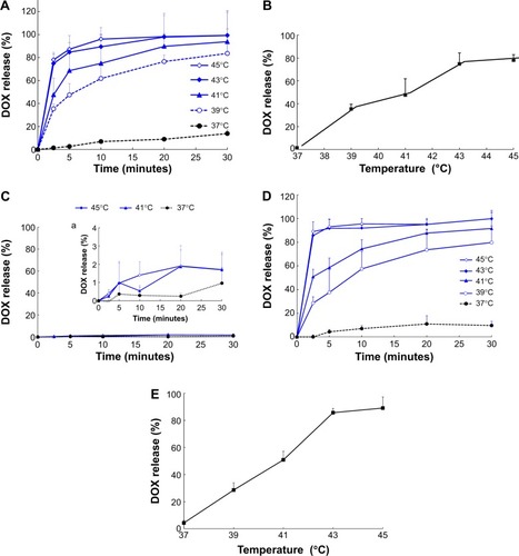
Table 2 The IC50 (μg/mL) values of DOX-LTSL-CREKA with or without heating at 43°C for 5 minutes in MCF-7/ADR and MCF-7 cells (n=3)
Figure 3 The flow cytometric measurement of DOX uptake from DOX-LTSL-CREKA by MCF-7/ADR cells.
Notes: Flow cytometric analyses (A) and mean fluorescence intensity (B) of MCF-7/ADR cells after 2-hour incubation with Roswell Park Memorial Institute 1640 medium (black line), free DOX unheated (red line), DOX-LTSL-CREKA unheated (green line), and DOX-SSL unheated (blue line) (a); free DOX heated at 43°C for 5 minutes before incubation (red fill), DOX-LTSL-CREKA heated at 43°C for 5 minutes before incubation (green fill), and DOX-SSL heated at 43°C for 5 minutes before incubation (blue fill) (b); free DOX heated at 43°C for 5 minutes after incubation (red grid line), DOX-LTSL-CREKA heated at 43°C for 5 minutes after incubation (green grid line), and DOX-SSL heated at 43°C for 5 minutes after incubation (blue grid line) (c); DOX-LTSL-CREKA unheated (green line), DOX-LTSL-CREKA heated at 43°C for 5 minutes before incubation (green fill), and DOX-LTSL-CREKA heated at 43°C for 5 minutes after incubation (green gird line) (d); DOX-SSL unheated (blue line), DOX-SSL heated at 43°C for 5 minutes before incubation (blue fill), and DOX-SSL heated at 43°C for 5 minutes after incubation (blue gird line) (e). **P<0.01 vs DOX-SSL; ##P<0.01 vs free DOX; &&P<0.01 vs unheated treatment; ‡‡P<0.01 vs heated before incubation treatment.
Abbreviations: DOX, doxorubicin; LTSL, lysolipid-containing thermosensitive liposome; CREKA, Cys-Arg-Glu-Lys-Ala; SSL, sterically stabilized liposomes.
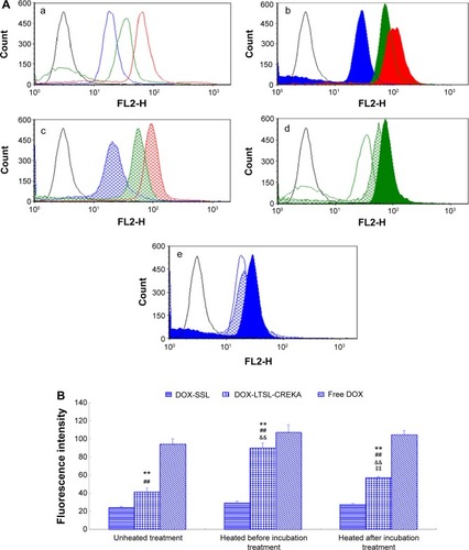
Figure 4 The confocal microscopy images of MCF-7/ADR cells incubated with DOX-LTSL-CREKA.
Notes: Confocal microscopy images of MCF-7/ADR cells incubated with DOX-LTSL-CREKA, DOX-SSL, and free DOX at unheated condition (a), with heating before incubation at 43°C for 5 minutes (b), and with heating after incubation at 43°C for 5 minutes (c) (A). Red is fluorescence of doxorubicin and blue is fluorescence of nuclei staining. Overlaying pictures of Hoechst 33258 and DOX are also shown (B) MCF-7/ADR cells incubated with DOX-LTSL-CREKA, DOX-SSL and free DOX at un-heated (a), with heating before incubation at 43°C for 5 minutes (b), with heating after incubation at 43°C for 5 minutes (c).
Abbreviations: DOX, doxorubicin; LTSL, lysolipid-containing thermosensitive liposome; CREKA, Cys-Arg-Glu-Lys-Ala; SSL, sterically stabilized liposomes.
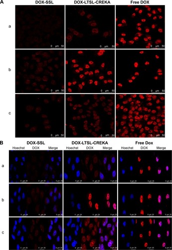
Figure 5 In vivo imaging.
Notes: The MCF-7/ADR tumor-bearing mice received an intravenous injection, via the tail vein, of physiological saline (A), cy7-LTSL (B), and cy7-LTSL-CREKA (C) at a dose of 750 μg/kg. Other tumor-bearing mice were intravenously injected heparin solution (a bolus of 800 units/kg), followed 120 minutes later by cy7-LTSL-CREKA (750 μg/kg), and received additional heparin by intraperitoneal injections (a total of 1,000 units/kg) throughout the experiment (D). All mice were then anesthetized with intraperitoneal injection of pentobarbital (60 mg/kg), and scanned at 2 hours, 6 hours, 8 hours, and 12 hours using a Kodak In Vivo Imaging System FX PRO.(p)(p)Abbreviations: cy7, carbocyanine dye; LTSL, lysolipid-containing thermosensitive liposome; CREKA, Cys-Arg-Glu-Lys-Ala.
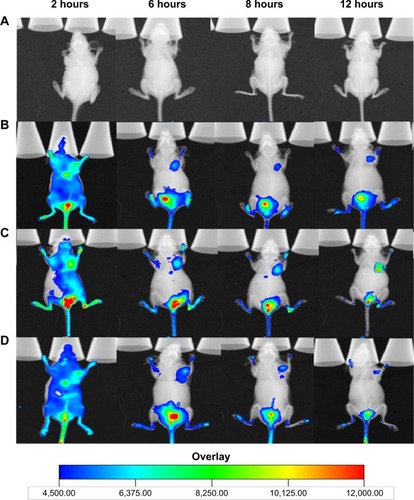
Figure 6 Localization of DiI-LTSL-CREKA in tumor tissue.
Notes: BALB/c nude mice bearing MCF-7/ADR tumors (~200 mm3) were intravenously injected with DiI-LTSL-CREKA at a dose of 200 μg/kg. At 2-hour or 6-hour time point, the mice in the experiment were sacrificed, the tumors were harvested, and tumor sections were examined for fluorescence. DiI-LTSL-CREKA (red) accumulates in tumor blood vessels (anti-CD31, green) (A) and co-localizes with anti-fibrin (ogen) (green) staining (B). The results are representative of four independent experiments. Magnification: ×200.
Abbreviations: DiI, 1,1′-dioctadecyl-3,3,3′,3′-tetramethylindocarbocyanine perchlorate; LTSL, lysolipid-containing thermosensitive liposome; CREKA, Cys-Arg-Glu-Lys-Ala.
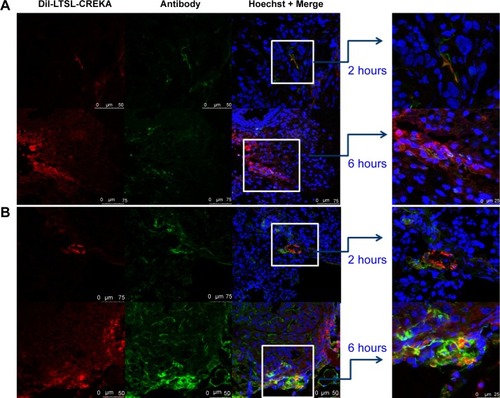
Figure 7 Tumor growth inhibition and tumor tissues histological analysis.
Notes: BALB/c mice were inoculated sc with MCF-7/ADR cells and treated with physiological saline, free DOX, DOX-SSL, DOX-LTSL, DOX-LTSL/Heated (tumor was locally heated at 43°C for 1 hour), DOX-LTSL-CREKA, and DOX-LTSL-CREKA/Heated (tumor was locally heated at 43°C for 1 hour) at the day 7 (4 mg/kg, iv, q2d ×3). Tumor size was measured with calipers twice per week. Results are given as means ± SD (n=6). (A, a) is the intact figure and (b) is the amplificatory figure. TUNEL staining of the paraffin-embedded tumors was performed according to the standard protocol provided by the manufacturers. Tumor apoptosis cells were detected by TUNEL (B). DNA strand breaks were labeled with (green), and nuclei were stained with Hoechst 332589 (blue). Apoptotic cells exhibited turquoise color as a result of color merge of these two labels. The fluorescence area of each group was used for the statistical analysis of apoptosis activity (C). **P<0.01 vs physiological saline as control group at the day 25. ##P<0.01 vs free DOX group at the day 25. $$P<0.01 vs DOX-LTSL or DOX-SSL group at the day 25. &&P<0.01 vs DOX-LTSL/Heated group at the day 25. ‡‡P<0.01 vs DOX-LTSL-CREKA group at the day 25.
Abbreviations: sc, subcutaneous; DOX, doxorubicin; SSL, sterically stabilized liposomes; LTSL, lysolipid-containing thermosensitive liposome; CREKA, Cys-Arg-Glu-Lys-Ala; iv, intravenous; SD, standard deviation; TUNEL, Terminal Deoxynucleotidyl Transferase Mediated dUTP Nick End Labeling.
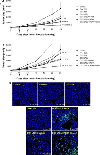
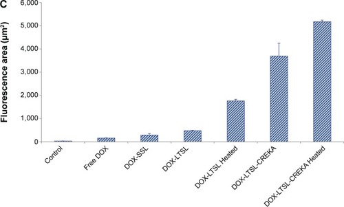
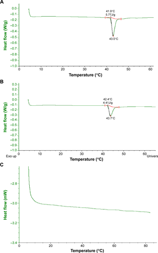
Figure S1 The DSC analysis of LTSL(A), LTSL-CREKA(B), and SSL(C).
Abbreviations: DSC, differential scanning calorimetry; LTSL, lysolipid-containing thermosensitive liposome; CREKA, Cys-Arg-Glu-Lys-Ala; SSL, sterically stabilized liposomes.