
Figures & data
Figure 1 Molecular design and characteristics of functionalized self-assembling peptide.
Notes: (A) Design and synthesis of KLD-F with FN and collagen IV mimic motifs, with KLD-F self-assembling into a nanoscale scaffold for encapsulating INS-1 β-cell. (B) Atomic force micrographs of KLD12 and KLD-F (0.1 mg/mL, bar 500 nm). (C) Width and height of KLD and KLD-F nanofibers (*P<0.05). (D) Photograph of KLD-F solution and hydrogel (10 mg/mL). (E) Rheological properties of KLD-F solution and hydrogel (Pa).
Abbreviations: ECM, extracellular matrix; FN, fibronectin.
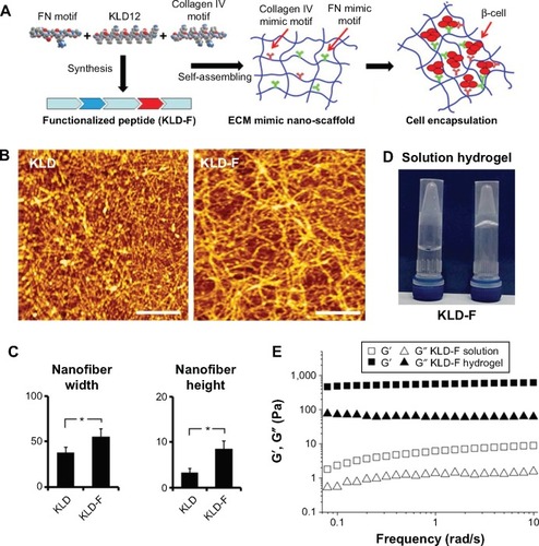
Figure 2 Functionalized self-assembling peptide enhanced ECM remodeling and cell-cell adhesion in INS-1 β-cells.
Notes: (A) Light and immunofluorescent micrographs of E-cad, FN, and Coll IV (bar 50 μm) after 3 days of culture. (B) Real-time polymerase chain reaction analysis of E-cad, FN, and Coll IV mRNA expression after 3 days of culture. (C) Western blot and quantitative analysis of E-cad and FN protein expression after 3 days of culture (*P<0.05).
Abbreviations: FN, fibronectin; E-cad, E-cadherin; Coll IV, collagen IV.
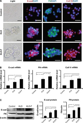
Figure 3 Functionalized self-assembling peptide improved insulin secretion function in INS-1 β-cells.
Notes: (A) Insulin secretion, (B) intracellular insulin (under 20 mM glucose), and (C) GSIS index in the different groups after 3 days of culture. (D) Real-time polymerase chain reaction analysis of Glut2, Ins1, MfaA, and Pdx-1 mRNA expression after 3 days of culture. (E) Immunofluorescent staining for insulin (under 20 mM glucose condition) and Pdx-1 (bar 50 μm). (F) Western blot and quantitative analysis of PDX-1 protein level after 3 days of culture (*P<0.05).
Abbreviations: DAPI, 4,6-diamidino-2-phenyllindile; GSIS, glucose-stimulated insulin secretion; Glu, glucose.
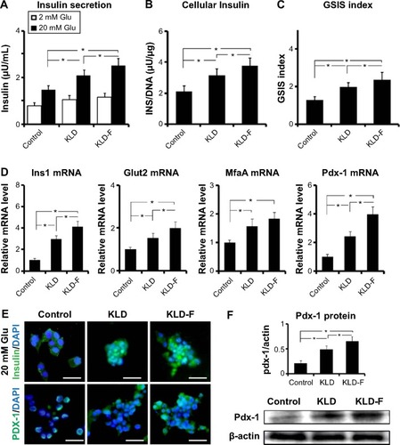
Figure 4 Functionalized self-assembling peptide promoted cell proliferation in INS-1 β-cells.
Notes: (A) Cell proliferation rates detected by CCK-8 assay after 3 days of culture. (B) BrdU enzyme-linked immunosorbent assay of INS-1 cell proliferation after 3 days of culture. (C) Glucose consumption rates (%) in different groups after 3 days of culture. (D) Western blot and quantitative analysis of Ki67 protein expression after 3 days of culture. (E) Immunofluorescent staining of Ki67 (bar 50 μm) and quantitative analysis of Ki67-positive cells (*P<0.05).
Abbreviations: CCK-8, Cell Counting Kit-8; DAPI, 4,6-diamidino-2-phenyllindile; ELISA, enzyme-linked immunosorbent assay.
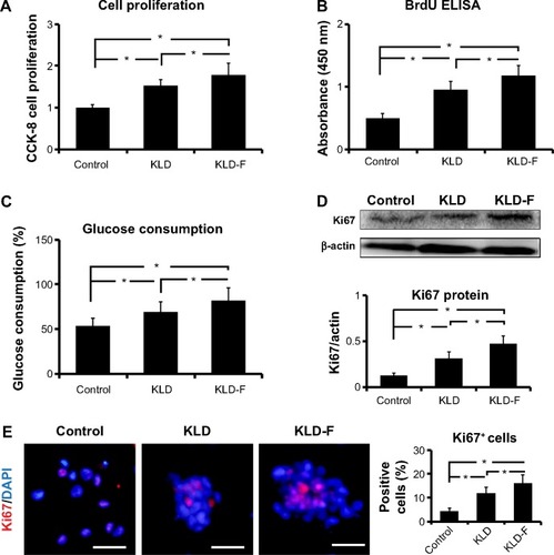
Figure 5 Functionalized self-assembling peptide induced cell cycle progression in INS-1 β-cells.
Notes: (A) Flow cytometry analysis of cell cycle. (B) G1, S, and G2/M phase distribution after 3 days of culture. (C) Proliferation index and (D) S-phase cell fraction detected by flow cytometry (*P<0.05). (E) Real-time polymerase chain reaction analysis of Ccnd1, Ccne1, Cdk2, and Cdkn1a mRNA levels after 3 days of culture (*P<0.05).
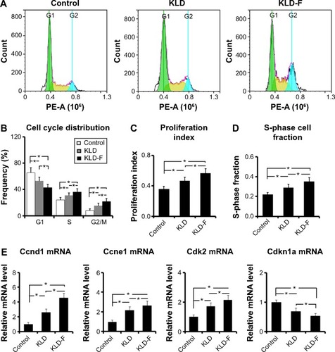
Figure 6 Mechanism of functionalized self-assembling peptide inducing β-cell proliferation.
Notes: (A–C) Western blot for p-FAK/FAK, p-Akt/Akt, and p-ERK/ERK after 1 day of culture, and quantitative analysis of protein level. (D–F) Western blot for integrin α5 and β1, cyclin D1, and p21 after 1 day of culture, and quantitative analysis of protein level (*P<0.05, **P<0.01). (G) Schematic map of signaling pathways. KLD-F binds to integrin α5 and β1, subsequently activating FAK/ERK and downstream cyclin D1 as well as inhibiting p27 signaling, finally promoting β-cell proliferation.
Abbreviations: ECM, extracellular matrix; FAK, focal adhesion kinase; ERK, extracellular signal-regulated kinase.
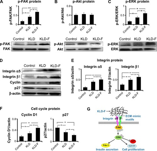
Table S1 Primer sequence for real-time polymerase chain reaction