Figures & data
Figure 1 X-ray diffraction patterns of TiO2 layers on quartz substrate.
Notes: (A) As grown and (B) annealed at 500°C.
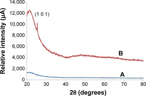
Figure 2 Field emission scanning electron micrographs of iron disk (A) and pin fixator (B).
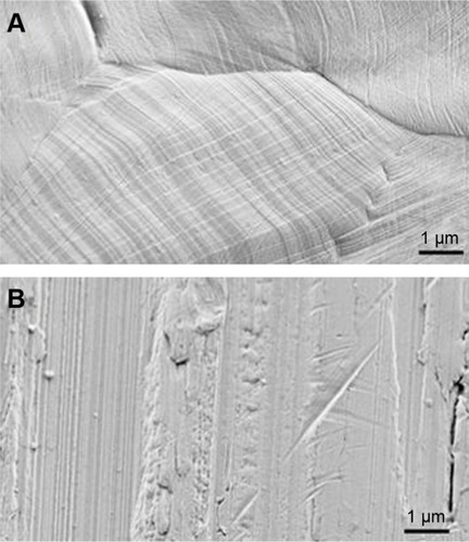
Figure 3 Field emission scanning electron micrograph of a punctual scratch on a cover layer.
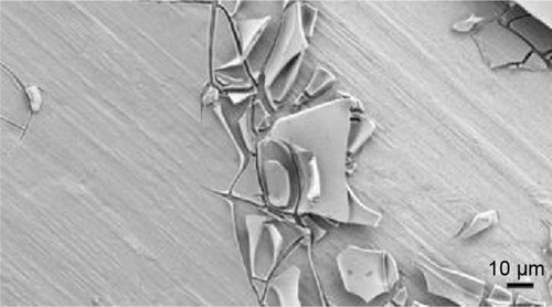
Figure 4 Field emission scanning electron micrograph showing the granular structure of the oxide layer at nanoscale.
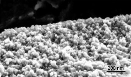
Figure 5 Illustrations of the mechanical test performed.
Notes: (A) Image of a unilateral fixator assembly performed on a cow femur. (B) Visualization of the insertion of the pin into the bone on a horizontal section.
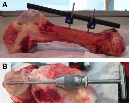
Figure 6 Field emission scanning electron micrograph of the contact area between the grip and the coated pin.
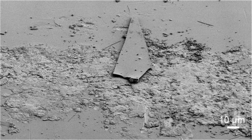
Figure 7 Field emission scanning electron micrograph of the contact area between bone and the coated pin after explantation.
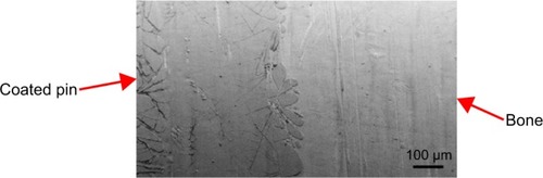
Figure 8 Photodegradation of AO7 solution in the presence of TiO2-coated disk versus UV irradiation time, as measured by the solution’s absorbance at λ =483 nm.
Abbreviations: AO7, acid orange 7; AU, absorbance units; UV, ultraviolet.
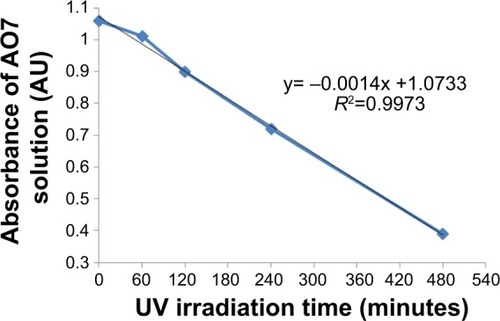
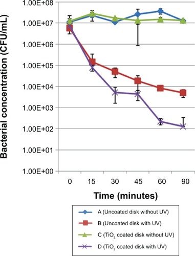
Figure 9 Inactivation kinetics for the Staphylococcus aureus strain. Comparison between coated and uncoated samples submitted to the same UV treatment duration.
Abbreviations: CFU, colony forming units; UV, ultraviolet.
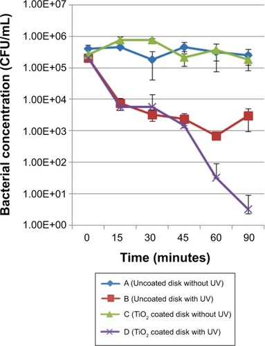
Figure 10 Inactivation kinetics for the Staphylococcus epidermidis strain. Comparison between coated and uncoated samples subjected to the same UV treatment duration.
Abbreviations: CFU, colony forming units; UV, ultraviolet.