Figures & data
Figure 1 The acoustic cavitation phenomenon that occurs under ultrasonic radiation and the NP creation.
Notes: The acoustic cavitation phenomenon happens when a bubble, which was created in the liquid, grows and then collapses. The bubble grows because the solute and/or the solvent vapors diffuse into the volume of the bubble, and it collapses when it gets to its maximum size. In the preparation of nano Penicillin and nano Vitamin B12, the material molecules are naturally drawn to the bubble surface, creating a shell of the material molecules around the bubble. During implosion, the molecules shell collapses into the bubble center and thereby creates a nanoparticle that contains many small molecules (a magnification of the nanoparticle is presented in the last phase of the process).
Abbreviation: NP, nanoparticle.
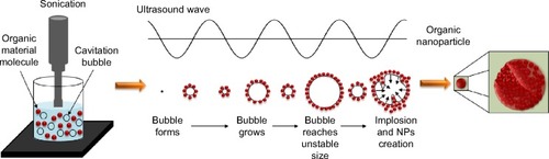
Figure 2 TEM images of Penicillin before and after the ultrasonic radiation.
Notes: (A) TEM image of Penicillin bulk form, (B) TEM image of Penicillin NPs after 5-minute sonication, (C) TEM images of Penicillin NPs after 10-minute sonication, (D) an enlargement of TEM image of Penicillin NPs after 10-minute sonication, and (E) diffraction lines of individual Penicillin nanocrystal.
Abbreviations: TEM, transmission electron microscopy; NPs, nanoparticles.
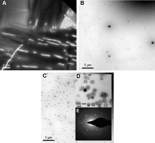
Table 1 Inhibition zone diameter for the different Penicillin NPs
Table 2 MIC for the different Penicillin NPs
Figure 3 Comparison of FTIR spectra of Penicillin before and after ultrasonic radiation.
Notes: FTIR spectrum of bulk Penicillin (solid line) and 5-minute ultrasonicated Penicillin (dashed line). The spectra show no change in the chemical structure of Penicillin upon 5-minute ultrasonic irradiation.
Abbreviations: FTIR, Fourier transform infrared spectroscopy; NPs, nanoparticles.
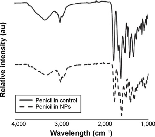
Figure 4 A comparison of size determination for Vitamin B12 by DLS measurements.
Notes: Size determination of bulk Vitamin B12 and Vitamin B12 upon 3- and 5-minute ultrasonic radiation.
Abbreviations: ONPs, organic nanoparticles; DLS, dynamic light scattering.
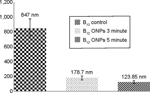
Figure 5 TEM images of Vitamin B12 before and after sonication.
Notes: (A) TEM image of Vitamin B12 bulk form. (B) TEM image of Vitamin B12 NPs upon 5-minute ultrasonic radiation.
Abbreviations: TEM, transmission electron microscopy; NPs, nanoparticles.
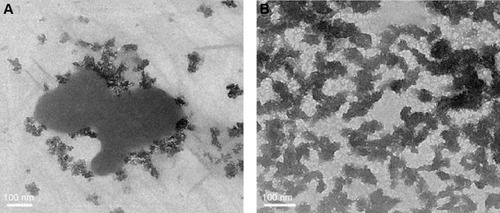
Figure 6 The absorption spectra of Vitamin B12 before and following ultrasonic radiation.
Notes: The absorption spectra of bulk Vitamin B12 (control, dashed line), Vitamin B12 following 3-minute sonication (3 minute, solid line), and Vitamin B12 after 5-minute sonication (5 minute, dots).
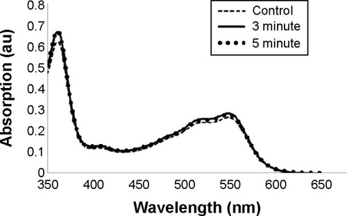
Figure 7 Hydroxyl radical formation via Fenton reaction in the presence of Vitamin B12 bulk form and NPs.
Notes: (A) The formation of hydroxyl radical via Fenton reaction. (B) The formation of hydroxyl radical in the presence of bulk Vitamin B12. (C) The formation of hydroxyl radical in the presence of nano Vitamin B12 obtained after 5-minute sonication.
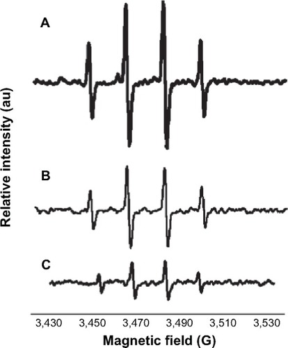
Figure 8 A comparison of hydroxyl radicals’ production.
Notes: The production of hydroxyl radicals (calculated by the integrated areas of DMPO spin adduct) via Fenton reaction in comparison with the production of hydroxyl radicals (calculated by the integrated areas of DMPO spin adduct) via Fenton reaction in the presence of bulk form and nano Vitamin B12 obtained after 5-minute sonication.
Abbreviation: DMPO, 5,5-dimethyl-1-pyrroline N-oxide.
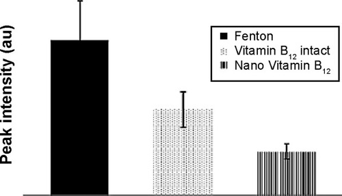
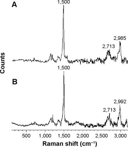
Figure 9 A comparison of Raman spectra of Vitamin B12 before and after ultrasonic radiation.
Notes: (A) Raman spectrum of bulk Vitamin B12. (B) Raman spectrum of Vitamin B12 upon 5-minute ultrasonic radiation. The results indicate the confirmation of the chemical structure of bulk form and nano Vitamin B12.