Figures & data
Figure 1 Synthetic route of 4-arm-PEG-TPGS.
Abbreviations: PEG, polyethylene glycol; TPGS, D-α-tocopherol polyethylene glycol succinate; SA, succinic anhydride; DMAP, 4-dimethylamino pyridine; DCC, dicyclohexylcarbodiimide; RT, room temperature.
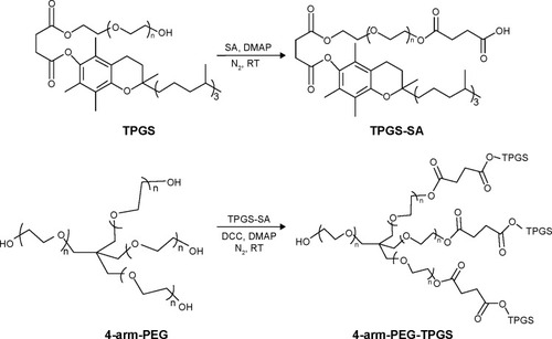
Figure 2 Characterization of 4-arm-PEG-TPGS.
Notes: (A) 1H-NMR spectra and (B) FTIR spectra.
Abbreviations: PEG, polyethylene glycol; TPGS, D-α-tocopherol polyethylene glycol succinate; NMR, nuclear magnetic resonance; FTIR, Fourier transform infrared spectroscopy; SA, succinic anhydride.
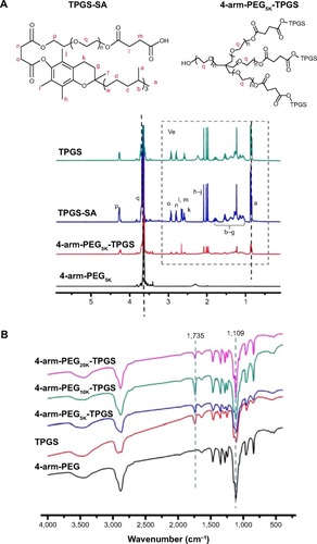
Figure 3 GPC results of 4-arm-PEG and TPGS-based derivative 4-arm-PEG-TPGS.
Abbreviations: GPC, gel permeation chromatography; PEG, polyethylene glycol; TPGS, D-α-tocopherol polyethylene glycol succinate; MW, molecular weight.
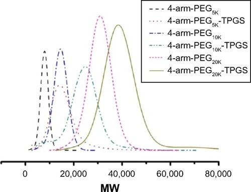
Table 1 Characterization of PTX-loaded 4-arm-PEG-TPGS nanoparticles
Figure 4 Characterization of 4-arm-PEG-TPGS nanoparticles.
Notes: (A and B) DLS result and TEM image of 4-arm-PEG5K-TPGS nanoparticles, (C and D) DLS result and TEM image of 4-arm-PEG10K-TPGS nanoparticles, (E and F) DLS result and TEM image of 4-arm-PEG20K-TPGS nanoparticles, (G) plot of the intensity ratio I339/I335 as a function of log C for TPGS, 4-arm-PEG5K-TPGS, 4-arm-PEG10K-TPGS, and 4-arm-PEG20K-TPGS nanoparticles, (H and I) the stability of TPGS micelles and PTX-NP dispersed in PBS and FBS, (J) hemolysis assay of blank 4-arm-PEG-TPGS nanoparticles of various concentrations incubated with RBCs for 4 hours at 37°C in an incubator shaker.
Abbreviations: PEG, polyethylene glycol; TPGS, D-α-tocopherol polyethylene glycol succinate; DLS, dynamic light scattering; TEM, transmission electron microscope; PTX, paclitaxel; NP, nanoparticles; PBS, phosphate buffered saline; FBS, fetal bovine serum; RBCs, red blood cells; PEI, polyethylenimine.
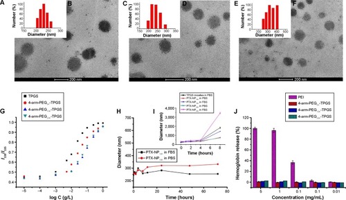
Figure 5 In vitro release of PTX from PTX-NP5K in PBS (pH 7.4) and FBS.
Abbreviations: PTX, paclitaxel; NP, nanoparticles; PBS, phosphate buffered saline; FBS, fetal bovine serum.
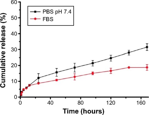
Figure 6 Cellular uptake of coumarin-6-NP5K by A2780 cells.
Notes: (A) CLSM images after 2 hours incubation and (B) MFI value analyzed by flow cytometry.
Abbreviations: NP, nanoparticles; CLSM, confocal laser scanning microscopy; MFI, mean fluorescence intensity; DAPI, 4′,6-diamidino-2-phenylindole.
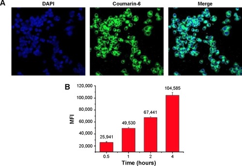
Figure 7 In vitro cytotoxicity of Taxol®, PTX-NP5K, and free PTX against (A) A2780, (B) A549, and (C) MCF-7 cells after treatment for 24, 48, and 72 hours.
Abbreviations: PTX, paclitaxel; NP, nanoparticles.
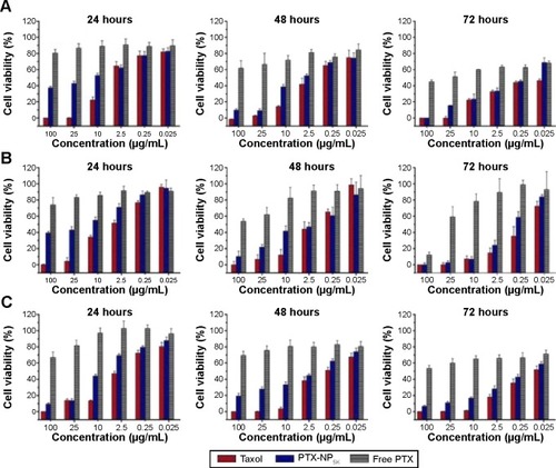
Table 2 IC50 values (μg/mL) of Taxol®, PTX-NP5K, and free PTX after 24, 48, and 72 hours incubation with A2780, A549, and MCF-7 cells at 37°C
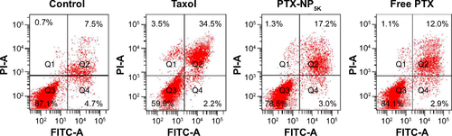
Figure 8 Cell apoptosis analysis of A2780 cells with Taxol®, PTX-NP5K, and free PTX after 24 hours treatment.
Notes: (A) Nucleus apoptosis assay and (B) annexin V-FITC/PI double staining by flow cytometry.
Abbreviations: PTX, paclitaxel; NP, nanoparticles; V-FITC, V-fluorescein isothiocyanate; PI, propidium iodide.
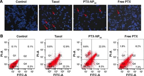
Figure 9 Cell cycle distribution in A2780 cells treated with various formulations.
Notes: (A) Control, (B) Taxol®, (C) PTX-NP5K, and (D) free PTX for 24 hours.
Abbreviations: PTX, paclitaxel; NP, nanoparticles; PI, propidium iodide.
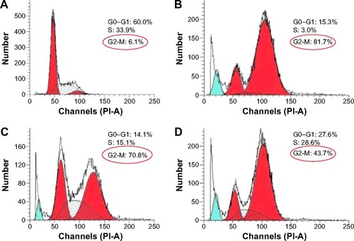
Figure 10 In vivo antitumor efficacy in tumor-bearing Kunming mice treated with Taxol® (10 mg/kg), PTX-NP5K (10 mg/kg), and PTX-NP5K (30 mg/kg) (n=5).
Notes: (A) Relative tumor growth ratio (*P,0.05), (B) tumor weight, (C) images of tumor tissues, (D) relative body weight and (E) HE staining assay of the tumor sections.
Abbreviations: PTX, paclitaxel; NP, nanoparticles; HE, hematoxylin and eosin.
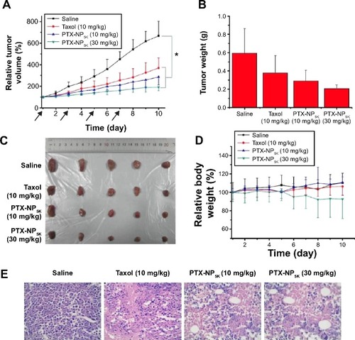
Figure S1 Hemolysis assay of Cremophor EL-based vehicle (Cremophor ELand dehydrated alcohol, 1:1, v/v) of various concentrations incubated with RBCs for 4 hours at 37°Cin an incubator shaker.
Abbreviation: RBCs, red blood cells.
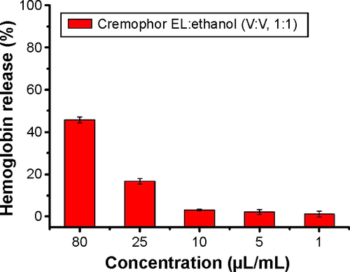
Figure S2 In vitro cytotoxicity of Taxol®, PTX-NP5K, and free PTX against S180 cells after treatment for 24, 48, and 72 hours.
Abbreviations: PTX, paclitaxel; NP, nanoparticles.
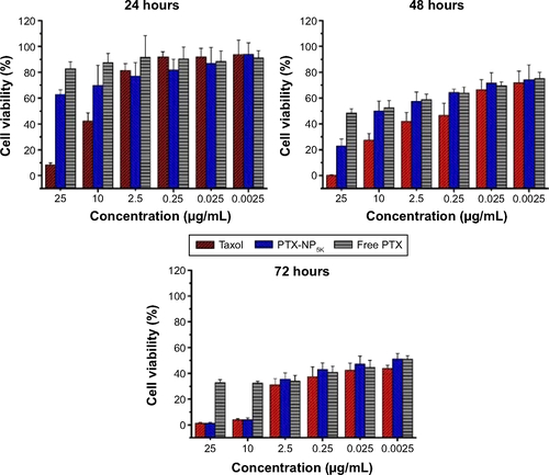
Figure S3 Cell apoptosis analysis of S180 cells with Taxol®, PTX-NP5K, and free PTX after 24 hours treatment.
Abbreviations: PTX, paclitaxel; NP, nanoparticles; FITC, fluorescein isothiocyanate; PI, propidium iodide.